Chapter 45 Diseases of the Reproductive System SNAKES Diseases of the reproductive system in the snake include dystocias, oviduct and cloacal prolapse, and penile prolapse. Some species of snakes such as pythons, king snakes, milk snakes, rat snakes, and corn snakes are oviparous (lay eggs), and others, including boas, vipers, and garter snakes are viviparous (live bearing). The testes of the male snake are found internally in the dorsomedial portion of the coelomic cavity. Snakes do not have an epididymis. The hemipenes are located in pouches lateral on each side of the cloaca. The location of the ovaries is similar to that of the testes. The oviducts have both an albumin and a shell-secreting function, and there is no true uterus. The oviducts empty into the cloaca. The onset of sexual maturity in snakes is affected by size, care, and diet more so than age. Snakes fed well and forced to grow rapidly can reach sexual maturity faster than snakes fed poor diets with slower growth rates. When raised under optimum conditions, many snakes will mature between 2 and 3 years of age. The majority of reptiles have a distinct breeding season, usually triggered by environmental stimuli such as light, temperature, rainfall, and the amount of food present. Many will begin breeding in the spring as temperatures warm; however, tropical snakes (the boas and pythons) tend to breed during the cooler periods of the season. The female must be in good health with energy reserves to breed; if not, breeding may not occur. A great deal of maternal energy is used for egg production or live births. Snakes that bear live young usually produce one clutch per year, whereas egg-laying snakes may produce a clutch several times. Female snakes may suspend feeding while giving birth and lose weight. Snakes provide no maternal care for their offspring except in choosing the site in which to deposit them. Neonates usually need no assistance in hatching and are ready to live on their own immediately after birth. Methods used to determine the sex of a snake include probing the pouches that contain the hemipenes (the deeper pouches are found in the male), by everting the hemipenes, or by observing the spurs found just lateral to the vent (larger in male snakes). Ultrasound or endoscopy can be used to determine the presence of ovaries in the female snake. Dystocia Dystocia is the most common reproductive problem seen in captive snakes. Dystocias may be divided into two groups: obstructive and nonobstructive. Obstructive dystocias result from an inability to pass one or more eggs or fetuses through the oviduct and cloaca. This may result from extra-large or malpositioned eggs or fetuses, from maternal abnormalities, or as a result of other masses obstructing the path. Nonobstructive causes may result from poor physical condition of the female snake, poor husbandry, improper nesting site, malnutrition, improper temperatures, and other environmental conditions. CLINICAL SIGNS • Visual presence of a mass in the cloaca or caudal abdomen • +/− Prolonged straining or cloacal prolapse • Often no clinical signs are obvious DIAGNOSIS • Physical examination and history • Ultrasound or radiographs TREATMENT • Physical manipulation to remove the retained egg or fetus • Hormonal stimulation; use of oxytocin or arginine vasotocin • Percutaneous ovocentesis—aspirate the contents of the egg through the ventrum of the snake to decrease the size of the egg • Surgery to remove retained eggs or fetuses • Supportive treatment of fluids, warmth INFORMATION FOR CLIENTS • Snakes must be kept at optimum conditions to facilitate reproduction. • Dystocia, although not a true emergency, should be corrected as soon as it is recognized. • Snakes with retained eggs may repeat the dystocia on future breeding. • Components of a good breeding program include two snakes of opposite sex, adult snakes in good health, optimal environmental conditions, and a good diet. Absence of one or more of these can affect the snake’s ability to reproduce. Oviduct/Cloaca (Colon) Prolapse Prolapse occurs with prolonged straining to pass eggs or fetuses, but it may also be caused by excessive straining for any reason (Fig. 45-1). With a shell gland/oviduct prolapse, the shell gland/oviduct will have a lumen but no feces will be present (as opposed to a prolapse of the colon with feces), and longitudinal striations appear on the surface of the shell duct that are not present on the colon. 
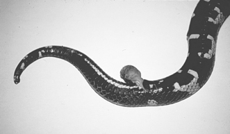
Figure 45-1 Prolapse of the hemipenis in a California Kingsnake. (From Mader DR: Reptile medicine and surgery, ed 2, St. Louis, 2006, Saunders, by permission.) CLINICAL SIGNS • Mass of reddish, moist, or dry tissue protruding from the vent • Straining • Lack of feces or urine elimination DIAGNOSIS • Physical examination and history of egg laying or straining TREATMENT • Gentle cleaning of the prolapsed tissue • Lubrication and manual reduction; purse-string suture of the vent may be required to temporarily maintain the reduction until swelling decreases Only gold members can continue reading. Log In or Register to continue You may also needDiseases of the Integumentary SystemDiseases of the Respiratory SystemDiseases of the Urinary SystemDiseases of the Musculoskeletal SystemDiseases of the Reproductive SystemDiseases of the Digestive SystemDiseases of the Urogenital SystemDiseases of the Endocrine System Share this:Click to share on Twitter (Opens in new window)Click to share on Facebook (Opens in new window)Click to share on Google+ (Opens in new window) Related Tags: Common Diseases of Companion Animals Aug 31, 2016 | Posted by admin in GENERAL | Comments Off on Diseases of the Reproductive System