Diseases of the Digestive System

Chapter 49 Diseases of the Digestive System


NORMAL DIGESTIVE SYSTEM ANATOMY


The digestive tract of the horse is that of an animal that was designed for grazing—that is, constant walking and eating. The lips of the horse, designed for prehension of grass, are amazingly sensitive and can sift out powdered medication when it is mixed in with their feed.


The horse’s teeth are that of an animal adapted for grazing. The teeth have enamel ridges that traverse the chewing surfaces and are constantly erupting throughout the horse’s life. As some of the enamel ridges get worn away, they are replaced by the ever-growing tooth. This constant tooth eruption, as well as the fact that the horse’s upper jaw is wider than its lower jaw, can lead to dental concerns as the horse goes through life.


The horse’s esophagus is a long, muscular tube that courses from the animal’s mouth to the stomach. The esophagus is mainly made up of two layers of smooth muscle: an inner circular layer and a longitudinal outer layer. These two layers of smooth muscle, and the way they work in concert, allow a horse to swallow its food.


The stomach of a horse is relatively small in comparison with its body size. The stomach of the average 1,100-pound horse has the same capacity of a 250-pound hog—roughly 2 to 4 gallons. There are two regions to the stomach: a nonglandular portion takes up roughly the proximal half, and the distal half is glandular. The small size of the stomach and its constant secretion of HCl have an impact on the horse’s feeding schedule, of which the owners need to be aware. The main function of the stomach is mixing of the feed with digestive juices and the start of protein digestion. The stomach also absorbs small amounts of water, alcohols, and water-soluble medications.


The small intestine is where most of the digestion and absorption of the concentrate portion of the feed occurs. Foodstuffs are broken down by chemical digestion mainly in the duodenum, which is the portion closest to the stomach. The resultant materials are absorbed as they pass through the jejunum and the ileum.


The fibrous portion of the diet empties from the ileum into the cecum. The cecum is a large, comma-shaped organ that comprises much of the right side of the horse’s abdominal cavity. The main function of the cecum is fiber digestion. Here, the microorganisms break down the fiber portion of the diet into glucose and volatile fatty acids (VFAs). The glucose is used by the gut microorganisms, and the VFAs are used by the horse for energy.


After digestion by the cecum, the remaining material goes into the large colon. The large colon has four portions: the right and left ventral and dorsal colons. The contents then travel into the transverse and then the descending colons. The main function of the colon is to absorb water from the feedstuff.


Many of the digestive system diseases that are seen in horses can be attributed to their somewhat peculiar digestive anatomy.


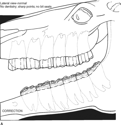
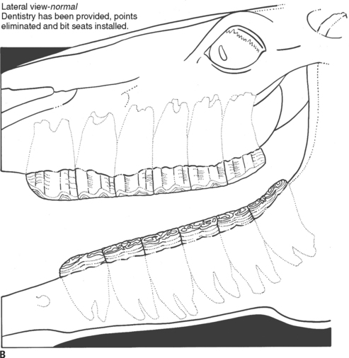
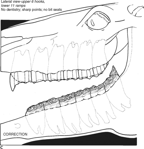
DENTAL CONSIDERATIONS: UNEVEN TOOTH WEAR


Because of the modification of diet and feeding patterns by domestication, demands on young performance horses, and the lack of selection for dental soundness, most horses need regular dental evaluation and work. Regular dental work can improve the horse’s comfort, increase efficiency of feed utilization, and possibly increase its performance.


As stated earlier, the horse’s teeth are constantly erupting throughout its life, and its upper jaw is wider than the lower jaw. This can lead to an uneven wear pattern of the occlusal surfaces of the teeth.


Common dental abnormalities that need attention include (Fig. 49-1):


CHOKE


Choke refers to a condition where a partial or total obstruction of the esophagus occurs, usually caused by feed impaction. Horses that choke often have a history of bolting their feed. Choke may also occur if a horse is fed too quickly after sedation. There are numerous potential causes for the esophagus to become obstructed. Causes of choke may include:


GASTRIC ULCERS


Gastric ulcers have become a much more frequently recognized disease in recent years. It is estimated that greater than 80% of show and race horses have some degree of mucosal ulceration.


The prevalence of gastric ulcers in horses is mainly due to management techniques. Most heavily worked horses are kept in stalls for 22 or more hours each day. They are fed high-concentrate meals rather than being allowed to graze freely all day. Feeding concentrate meals predisposes a horse to ulcers by two major pathways:


Saliva contains bicarbonate and acts as a natural buffer to the stomach acids. A horse can salivate only when it has something in its mouth. A grazing horse is salivating nearly constantly, whereas one fed in meals salivates only at mealtime. A grazing horse also has a constant physical buffer in the stomach to protect it from the acid environment. Concentrate feeds also contribute to the problem because concentrates produce one-fourth the amount of saliva when compared with an equal amount of forages.


NSAIDs such as flunixin meglumine and phenylbutazone have been shown to cause ulcers in the stomach and other portions of the digestive tract, especially in the right dorsal colon. If possible, the use of NSAIDs should be limited to 5 to 7 days. Oral NSAIDs should also be administered with a meal.


Aug 31, 2016 | Posted by in GENERAL | Comments Off on Diseases of the Digestive System

Full access? Get Clinical Tree

Get Clinical Tree app for offline access