CHAPTER | 13 Diseases of Eyes, Claws, Anal Sacs, and Ear Canals


Blepharitis
Features
Insect Bite/Sting Hypersensitivity
Onset of eyelid erythema, with swelling (angioedema) or raised focal masses, is acute.
Diagnosis
Treatment and Prognosis


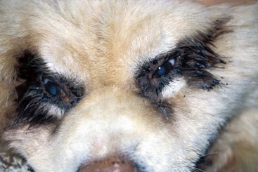
FIGURE 13-1 Blepharitis.
Discoloration and matting of the hair around the eye in a dog with bilateral blepharitis.
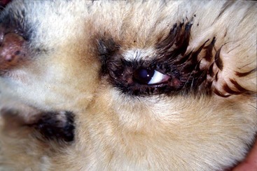
FIGURE 13-2 Blepharitis.
Close-up of the dog in Figure 13-1. Discoloration and matting of the hair around the eye caused by the copious ocular discharge are apparent.
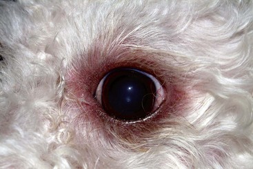
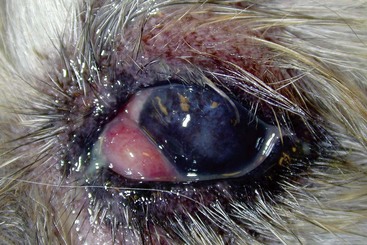
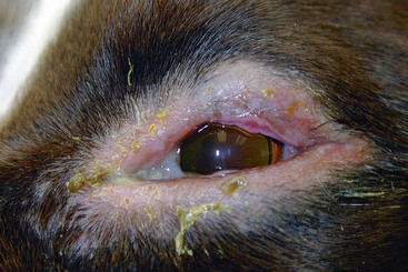
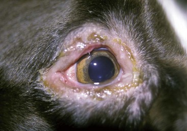
FIGURE 13-6 Blepharitis.
Same dog as in Figure 13-5. The “marginal blepharitis” caused a bilateral, alopecic, papular dermatitis.
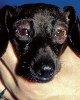
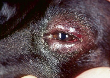
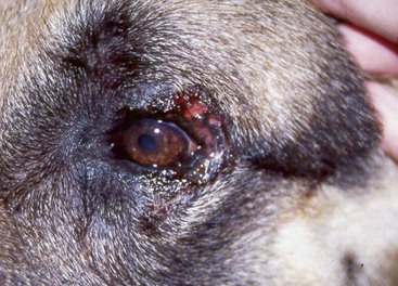
FIGURE 13-8 Blepharitis.
Same dog as in Figure 13-7. Alopecia, erythema, and tissue swelling of the periocular skin.
(Courtesy S. McLaughlin.)
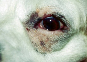
FIGURE 13-9 Blepharitis.
Alopecia and erythema affecting the periocular skin caused by a secondary bacterial pyoderma.
(Courtesy E. Willis.)
Otitis Externa
Features
Otitis externa is an acute or chronic inflammatory disease of the external ear canal. Its causes are numerous and almost always have an underlying primary disease (Table 13-1) that alters the normal structure and function of the canal, resulting in a secondary infection (Table 13-2). Otitis externa is common in cats and dogs, with Cocker spaniels especially at risk for developing severe and chronic disease.
TABLE 13-1 Primary Causes of Otitis Externa
| Primary Factor | Characteristics | Comments |
|---|---|---|
| Parasites | Otodectes cynotis | Cause approximately 50% of otitis cases in cats and 5% to 10% of otitis in dogs. Dogs and cats can be asymptomatic carriers |
| Demodicosis | Can cause a ceruminous otitis in dogs and cats | |
| Sarcoptes scabiei | Typically, the ear margin and ventral third of the outer ear pinna are affected. Otitis externa is not usually a feature of this disease | |
| Hard ticks, chiggers | May affect ear pinnae and external ear canal | |
| Spinous ear ticks | An uncommon cause of otitis externa in dogs and cats | |
| Hypersensitivities | Atopy | Otitis externa is seen in 50% to 80% of atopic dogs. (In 3%–5% of them, otitis externa is the only symptom.) Usually, bilateral otitis |
| Food hypersensitivity | Otitis externa is seen in up to 80% of dogs with food hypersensitivities. (In more than 20% of these dogs, otitis externa is the only symptom.) | |
| Contact dermatitis | Otic medication (e.g., neomycin, propylene glycol) can cause irritant reactions in the ear. Should be suspected any time ear disease worsens significantly while animal is undergoing topical treatment | |
| Endocrine Disorders | Hypothyroidism | Bilateral ceruminous otitis externa. Most common in middle-aged to older dogs. Usually, skin involvement |
| Foreign Bodies | Usually present as unilateral otitis externa. Look for plant material, dirt, small stones, impacted wax, loose hair, and dried medication. Often, the inciting foreign body is not identified because it becomes so coated with cerumen that, when removed during ear flushing, it is not recognizable | |
| Keratinization Disorders | Canine primary seborrhea | Bilateral ceruminous otitis. Usually have other skin involvement, especially Cocker spaniels |
| Facial dermatosis of Persians | Bilateral ceruminous otitis externa and seborrheic facial dermatitis. Secondary malasseziasis is common. Uncommon to rare in Persian cats | |
| Sebaceous adenitis | May cause dry, scaly ears and mild inflammation. Usually other skin involvement. Rare in dogs, with highest incidence in Standard Poodles, Akitas, and Samoyeds | |
| Autoimmune/Immune-Mediated Diseases | Juvenile cellulitis | Acute cellulitis of muzzle and periocular regions with marked submandibular and prescapular lymphadenomegaly. Exudative otitis externa, fever, and depression may also be present. Uncommon in puppies 3 weeks to 6 months old, with highest incidence in Golden retrievers, Labrador retrievers, Dachshunds, Pointers, and Lhasa apsos |
| Inflammatory Polyps (cats) | May present as recurrent, unilateral otitis externa. Polyps may originate from lining of tympanic cavity, auditory canal, or nasopharynx | |
| Neoplasia | Cats | Ceruminous gland adenomas and adenocarcinomas, sebaceous gland adenomas and carcinomas, squamous cell carcinomas, papillomas |
| Dogs | Ceruminous gland adenomas and adenocarcinomas, papillomas, basal cell carcinomas, squamous cell carcinomas | |
| Conformation | Heavy, pendulous ears | May result in decreased air circulation, increased heat, and moisture |
| Narrow ear canals Retention in the ear canal | Nidus for infection | |
| Hair in ear canals | ||
| Increased glandular tissue |
TABLE 13-2 Secondary Causes of Otitis Externa
| Secondary Factors | Comments |
|---|---|
| Bacterial infection | Include Staphylococcus spp., Streptococcus, Pseudomonas spp., Proteus, and Escherichia coli. Recurrent bacterial otitis is often associated with underlying allergies. |
| Yeast infection | Malassezia pachydermatis. Recurrent yeast otitis is often associated with underlying allergies. |
| Otitis media | Chronic otitis externa (2 months’ duration or longer) often results in extension of the disease into the middle ear. The otitis media can then be a source for recurrent otitis externa. |
| Chronic pathologic changes | With chronic inflammation, the dermis and the subcutis become fibrotic, leading to permanent stenosis of the canal lumen. The auditory cartilage may become calcified and ossified. Secretions, desquamated cells, and proliferating microorganisms become entrapped. Calcified ear cartilage is a permanent change that cannot be resolved with medical therapy. |
Diagnosis
Treatment and Prognosis
Products that can be used for ear flushing include the following:


Individual Diseases
 Otic miticide as per label directions (ivermectin and milbemycin products are safe and highly effective)


 dropperful) should be instilled in the affected ear every 12 hours for at least 2 to 4 weeks. Treatment should be continued until follow-up ear smears are cytologically negative for microorganisms, the external canals are no longer edematous or inflamed, and the ear canal epithelium has normalized.
Effective products include the following:


 dropperful) should be instilled in affected ears every 8 to 12 hours for at least 2 to 4 weeks. Treatment should be continued until follow-up ear smears are cytologically negative for microorganisms, the external canals are no longer edematous or inflamed, and the ear canal epithelium has normalized.
Effective products include the following:


Antibiotics include the following:


 The EDTA solution should be combined with enrofloxacin to make a 10- to 20-mg/mL solution. The solution should be used q 12–24 hours to completely fill the ear canal. Even as the sole therapy, the surfactants in T8 Solution act to clean the ear while allowing the high concentration of enrofloxacin to penetrate into the deep canal. This treatment is 80% effective in chronic, recurrent otitis cases, even if the bacteria are reported to be resistant to enrofloxacin (because of the tris-EDTA and high concentration of antibiotic).


 tsp]) of Silvadene Cream with 13.5 mL distilled water, or mix 0.1 g silver sulfadiazine powder with 100 mL distilled water), and instill 0.5 mL q 12 hours

For End-Stage Ears


