Fig. 46.1
Light micrographs of serial paraffin sections prepared by IVCT and stained with H&E (HE, a–c) or immunostained for albumin (d–f), immunoglobulin G 1 (IgG1 , g–i), or immunoglobulin M (IgM , j–l). Note different immunostaining intensities of serum protein s in the interstitium between tumor cells (e, h, k). In the tumor cell nests (e, h, k), albumin and IgG1 are more obviously immunolocalized (black arrows, e and h), as compared to IgM immunostaining (black arrows, k). (f, i, l) Immunoreactivities of albumin, IgG1, and IgM are detected in the interstitium between tumor cells (black arrows). (d–l) All three serum proteins are also immunolocalized in blood vessel s (arrowheads) and connective tissues around the tumor mass (white asterisks), but different intensities of immunoreactivities for three proteins are seen in connective tissues invading beyond the tumor capsule (white arrows). Black asterisks, glandular organization. Bars 50 μm
46.3 Immunodistribution of Extrinsic BSA in Xenografted Lung Adenocarcinoma Tissue
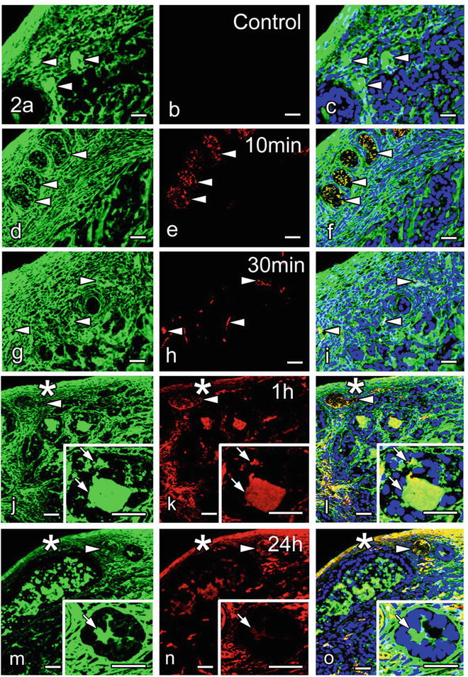
The examination of the injected BSA is important to know how soluble serum components pass through blood vessel s and diffuse into the tumor connective tissues. BSA was mostly immunolocalized within blood vessels in 10 and 30 min after injection (see Fig. 46.2d–i), but it was diffusely immunolocalized throughout the tumor interstitium in 1 h. Our findings in the present study were nearly in agreement with some previous data shown by live imaging with intravital or confocal laser scanning microscopy [8], indicating that the BSA leaked out into the interstitial spaces quickly, within 1 h. Although another study reported that no difference of molecular leakage was seen among the different types of tumors [3], the relative rate of BSA leakage into the tumor interstitium might be significantly changed within the tumor tissues, which would partly depend on the different molecular structures of interstitial matrices and functions of blood vessels [9, 10] or variously elevated interstitial fluid pressures [11, 12]. This morphofunctional discrepancy in the tumor tissues was directly visualized by the injected BSA in living mice.


Fig. 46.2


Double-immunofluorescence micrographs of albumin (green) and intravenously injected bovine serum albumin (BSA) (red) with nuclear staining (Topro3; blue) in cryosections prepared by IVCT: control (a–c), and 10 min (d–f), 30 min (g–i), 1 h (j–l), or 24 h (m–o) after the BSA injection. Arrowheads indicate blood vessel s . (d–i) In 10 and 30 min, BSA immunoreactivity is seen only in blood vessels (arrowheads). (j–o) In 1 and 24 h, however, it is also detected in the extracellular space s between tumor cells (small arrows in insets) and connective tissues around the tumor mass (asterisks). Bars 60 μm
Stay updated, free articles. Join our Telegram channel

Full access? Get Clinical Tree


