Differential Diagnoses

CHAPTER | 1 Differential Diagnoses


Almost all dermatology patients have a primary/underlying disease that causes secondary infections. These infections must be eliminated and prevented but will recur rapidly unless the primary disease is identified and controlled.


Most skin cases seen in a veterinary practice can be successfully managed if two essential questions can be answered: (1) What are the secondary infections? (2) Why are these secondary infections there?


What Are the Infections?


A vast majority of dogs with allergy or endocrine disease have or will have a secondary bacterial or yeast infection. Yeast dermatitis is the most commonly missed diagnosis in general practice dermatology. Bacterial pyoderma is often identified but is usually mistreated with too low doses of antibiotics administered for too short a time. Otitis is now recognized and treated better than it was in years past; however, treatment for otitis that is based on actual documented organism types and relative counts on follow-up evaluations is a rare occurrence.


So, what is the solution?


For every dermatitis case every time you evaluate the patient, ask yourself, “What are the infections?”


Unless you have microscopic vision, answering this question will require the use of cytology. Unfortunately, most general practices do not routinely perform skin and ear cytology for dermatitis; instead they rely on the doctor’s best guess. Sometimes this can be successful (even a broken clock is correct twice a day); however, a more precise method is available. Use of diarrhea and the fecal exam as a comparison and as a model for improvement works well because both skin cytology and fecal exams involve the use of a microscope, can easily identify the type of infection, and can be performed by trained technical staff.


The answers to these questions should be the same for skin cytology (skin scrapings, impression smears, tape preps, and otic swabs).


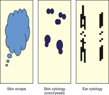
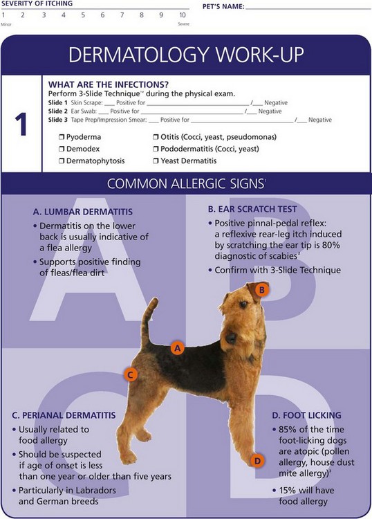
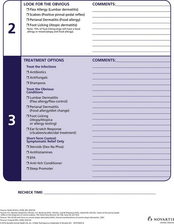
The practical solution for determining the best method by which to answer the question, “What are the infections?” is to implement a minimum database infection screening procedure to be performed by the technician before the veterinarian examines the patient. Every dermatology patient should undergo otic cytology, skin cytology (an impression smear or a tape prep), and a skin scrape at every examination (initially and at every recheck visit). The 3-slide technique (Figure 1-1) can be performed easily and interpreted by a technician before the doctor completes an evaluation—which is exactly how diarrhea and fecal examinations are handled in most clinics. Moving the cytologic evaluation to the beginning of the dermatology appointment and thereby empowering the technical staff to accomplish the evaluation optimizes the dermatology appointment and provides essential information in the most efficient manner.


When an owner brings a pet into the clinic for a small hairless spot, it would be appropriate to question the necessity for an otic cytology when the hairless spot is the problem. However, the 3-slide technique is most helpful in these exact types of cases. If focal pruritus occurs in a dog and the patient has a secondary otitis (which the technician identified during the infection screen), the veterinarian should more aggressively discuss this and work up the patient for possible allergy. If the patient did not have otitis, the pruritus could be minimized in the hope that it was a short-term problem that is likely to self-resolve.


Similarly, there is no excuse for mistreating a patient who has demodicosis. Lesions caused by demodicosis can look identical to folliculitis lesions caused by bacterial pyoderma and dermatophytosis. Clinical appearance is not an acceptable criterion for ruling in or ruling out demodicosis. When the technician performs a skin scrape as part of the infection screen, demodicosis can be identified and treated easily and accurately.


Yeast dermatitis is one of the most frequently missed diagnoses in the United States as of this writing. The clinical presentation is unique; however, initial cytologic evaluation is often omitted, and follow-up cytology to determine the effectiveness of treatment rarely happens.


Why Are They There?


Infections are always secondary to a primary disease; however, all too often, the patient is not evaluated or treated for the primary disease for three main reasons: (1) only the secondary infections are treated over and over again, (2) the nature of the allergy is confusing, and (3) cheap steroids that have delayed repercussions are accessible.


Why are the infections there? This question should be asked and answered for every dermatology patient if successful outcomes are to be achieved.


Most dermatology patients suffer from allergy or endocrine disease. Through signalment, a good patient history, and recognition of unique patterns of lesions, a prioritized differential list can be formulated quickly.


By knowing the most unique and frequent symptoms associated with each allergic disease, an astute clinician can determine the most likely allergy with approximately 85% accuracy; this rate rivals many other diagnostic testing results for some of the most common assays.


For example, a dog that is foot licking is likely atopic. If the owner reports a seasonal pattern to the podopruritus, then you have a reasonably accurate diagnosis—EASY.


Author’s Note


Sep 10, 2016 | Posted by in SMALL ANIMAL | Comments Off on Differential Diagnoses

Full access? Get Clinical Tree

Get Clinical Tree app for offline access