CHAPTER 110 Diagnostic Imaging and Surgical Management of Conditions Involving the Stifle Joint
The equine stifle joint is anatomically complex and technically challenging to evaluate fully because of the large size of the joint itself and the surrounding muscle mass. Isolation of lameness to the stifle joint is typically based on a thorough physical examination, including palpation of accessible anatomic structures: the three joint compartments (femoropatellar, medial femorotibial, and lateral femorotibial), patella, distal patellar ligaments, and collateral ligaments. In addition, a thorough lameness examination with intra-articular anesthesia of all three joint compartments is frequently necessary to localize lameness to the stifle joint.
IMAGING MODALITIES
Radiography
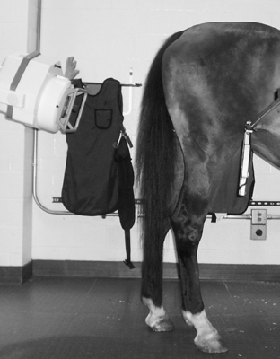
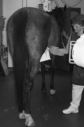
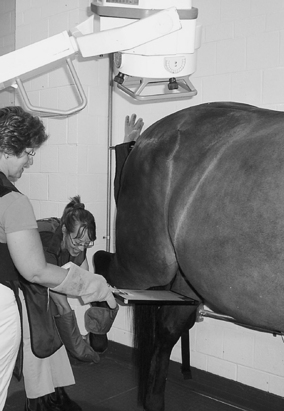
Radiography is typically the first imaging method used to evaluate the bony and soft tissue structures of the stifle joint. When possible, images should be obtained using digital radiography because they have significantly better contrast, elucidation of bone structure, and diagnostic value compared with images obtained using conventional radiography. A minimum of three survey projections should initially be used to obtain images, including caudocranial (with x-ray tube angled 0 to 15 degrees distad, depending on the conformation of the horse; Figure 110-1), lateromedial, and caudal 60 degrees lateral-craniomedial oblique views (Figure 110-2). A detailed map of the soft tissue attachments that includes tendons, ligaments, and fibrous portion of the joint capsules has been drawn for these three standard projections, and they can be useful when interpreting stifle radiographs. Only the caudolateral to craniomedial oblique view (and not a caudomedial or craniolateral oblique) is included in the survey set because the medial femoral condyle and the lateral trochlear ridge of the femur, which are often affected in the horse, are projected. This projection can provide important information about the depth of an osteochondritis desiccans (OCD) lesion of the femoral lateral trochlear ridge or the configuration of a medial femoral condyle subchondral bone cyst. When indicated, additional views, including a caudal 60-degree medial-craniolateral projection, flexed lateromedial, and skyline projection of the patella (Figure 110-3), should be obtained.
Magnetic Resonance Imaging
Assessment of the musculoskeletal system using magnetic resonance imaging (MRI) has advanced considerably in recent years. Although not widely available for imaging the equine stifle, MRI is the most effective imaging modality for assessing the entire joint, including cartilage, meniscus, ligaments, and subchondral bone sclerosis and edema (Figure 110-4
Stay updated, free articles. Join our Telegram channel

Full access? Get Clinical Tree


