Fig. 9.1
Light microscopic images of HE staining (a, d, g, j, m, p, s, v) and immunostaining of ERK1/2 (b, e, h, k, n, q, t, w) and phosphorylated ERK1/2 (c, f, i, l, o, r, u, x) on serial paraffin sections in mouse small intestine with different preparative methods, such as IVCT-FS (a–f), QF-FS (g–l), IM-DH (m–r), and PF-DH (s–x), in crypts (a–c, g–i, m–o, s–u) and villi (d–f, j–l, p–r, v–x). Arrowheads (c, f, i, l, r) show the immunopositive staining of pERK1/2 in the crypts and villi. ML muscular layer, Cr crypt, and Vi villi. Bars, 20 μm
By IM-DH, congested erythrocytes and shrinkage of cells and tissues were observed upon HE staining (Fig. 9.1m, p). ERK1/2 immunoreaction was detected in both cytoplasm and nuclei of cells in all mucous layers (Fig. 9.1n, q). However, pERK1/2 was detected only in epithelial cell s at the top parts of villi , but not in the crypt areas (Fig. 9.1o, r). By PF-DH, erythrocytes were washed out during perfusion fixation ; the cytoplasm of epithelial cells was strongly stained with eosin, and the total cell volumes and their nuclei had shrunk (Fig. 9.1s, v). ERK1/2 immunoreaction was detected mainly in the nuclei of epithelial cells of all layers (Fig. 9.1t, w), but pERK1/2 was hardly detected in those areas (Fig. 9.1u, x). Thus, different immunolocalizations of ERK1/2 and pERK1/2 were demonstrated by the different preparative methods. In particular, pERK was hardly detected with IM-DH or PF-DH. We assumed that IVCT-FS more clearly revealed morphological images and immunolocalization of pERKs, mostly reflecting their living state s , with less effect of ischemia or technical diffusion artifact s .
9.3 ERK1/2 and pERK1/2 in 20 min Ischemia Group with IVCT-FS and IM-DH
Using morphological images with HE staining (Fig. 9.2a), many erythrocytes were shown to be congested in blood vessel s , and some epithelial cell s at the top parts of villi were swollen due to ischemia (Fig. 9.2a, d, g, j). The immunolocalization of ERK1/2 was seen in the cytoplasm of epithelial cells (Fig. 9.2b, e, h, k), which was almost the same as that of the sham operation group. pERK1/2 immunostaining was clearly detected in the cytoplasm and nuclei of epithelial cells in the crypt (arrowheads in Fig. 9.2f, l), but not in epithelial cells at the top parts of villi (Fig. 9.2i). Such immunopositive staining of both ERK1/2 and pERK1/2 was confirmed by performing IVCT from the luminal sides (the inset in Fig. 9.2b and the left inset in Fig. 9.2c), by detecting the immunostaining of ERK1/2 in the cytoplasm of epithelial cells and pERK1/2 in the epithelial cells at the crypt areas. Immunostaining patterns of ERK1/2 and pERK1/2 were slightly different with another IM-DH preparation under the 20 min ischemia conditions (insets in Fig. 9.2d–i). Intestinal tissues were mostly shrunken due to alcohol dehydration during the conventional tissue preparation (insets in Fig. 9.2d, g). ERK1/2 was immunolocalized in both cytoplasm and nuclei of cells in all layers (insets in Fig. 9.2e, h). The immunolocalization of pERK1/2 was weakly detected in the nuclei of epithelial cells at crypt areas, compared with the immunostaining of pERK1/2 with IVCT after 20 min ischemia, not at the top areas of villi (insets in Fig. 9.2f, i). These findings indicate that the ERK1/2 signal transduction was usually activated in the intestinal epithelial stem cells under ischemia for 20 min.
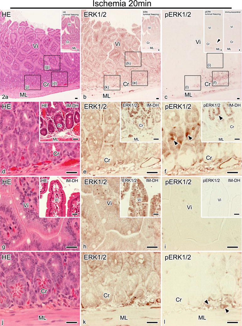
Fig. 9.2


Light microscopic images of HE staining (a, d, g, j) and immunostaining of ERK1/2 (b, e, h, k) and pERK1/2 (c, f, i, l) on serial paraffin sections in living mouse small intestines with IVCT and IM-DH under the 20 min ischemic conditions. Inset in (a): HE staining by preparing samples with freezing from luminal sides. Inset in (b): immunostaining of ERK1/2 by preparing samples with freezing from the luminal sides. Left inset in (c): immunostaining of pERK1/2 by preparing samples with freezing from the luminal sides. Right inset in (c): immunocontrol with a secondary antibody alone. Inset in (d–i): HE staining and immunostaining of ERK1/2 and pERK1/2 on serial paraffin sections in mouse small intestines with IM-DH. (d–l): highly magnified image of rectangles shown in the top panel. Arrowheads in (f, l) show the immunopositive staining of pERK1/2 in epithelial cell s of crypts. ML muscular layer, Cr crypt, and Vi villi. Bars, 20 μm
Stay updated, free articles. Join our Telegram channel
Full access? Get Clinical Tree
