9 Dermatophytosis in a Jack Russell terrier
CASE HISTORY
The relevant aspects of the history were:
• New lesions consisting of alopecia, erythema, crusting and scaling had subsequently appeared over the face, limbs and tail.
• Slight weight loss had been noted over the past month, but the dog had remained bright and active.
• One other dog and a cat were part of the household. The dog had developed a similar lesion over the bridge of the nose, although this had resolved spontaneously.
• Previous treatment with several weekly amitraz dips at a concentration of 500 ppm and various 7- to 10-day courses of systemic antibacterial therapy had not resulted in any clinical improvement.
CLINICAL EXAMINATION
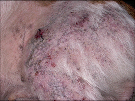
Examination of the skin revealed extensive patches of relatively well-demarcated alopecia, erythema, hyperpigmentation, and crusting and scaling over the muzzle, periorbital skin, the left dorsal trunk, the lateral thighs and the forelimbs (Figs 9.1, 9.2 and 9.3).
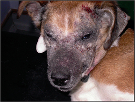
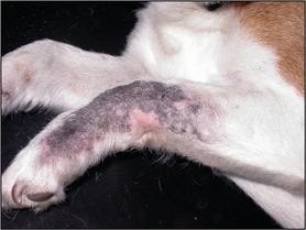
Figure 9.2 Well-demarcated patch of alopecia, erythema and hyperpigmentation over the left carpal area.
CASE WORK-UP
The following diagnostic tests were performed:
• Microscopic examination of multiple deep skin scrapings, which failed to reveal the presence of Demodex spp. mites.
• Cytological examination of stained impression smears from the underside of the crusts (Fig. 9.4), which revealed degenerate neutrophils and small numbers of intra- and extracellular cocci. Small numbers of acantholytic keratinocytes were also evident (Fig. 9.5).
• Fungal culture on material collected using the MacKenzie brush technique, together with plucked hairs and scrapes, which grew Trichophyton erinaciei.
Stay updated, free articles. Join our Telegram channel

Full access? Get Clinical Tree


