28 Dermatophytosis
CLINICAL EXAMINATION
Feline dermatophytosis can present not only with a variety of different lesions, but also with different reaction patterns, which are all common to other diseases seen in this species. The alopecia may be circumscribed or diffuse, with patches of scaling, hyperpigmentation, erythema and comedone formation, which may be present on the head or on the extremities. The hairs on the margin of lesions may be broken. In some feline cases clinical reaction patterns such as miliary dermatitis, eosinophilic granuloma complex and chin acne have been associated with dermatophyte infection. Dermatophyte pseudomycetoma, a form seen in long-haired cats, is characterized by subcutaneous nodules, which may ulcerate and/or discharge.
In dogs the clinical signs, although variable, are mainly associated with hair loss. The lesion distribution may be localized or diffuse. Circular patches of alopecia, scaling, hyperpigmentation, crusts and/or follicular papules may be seen on the head and the extremities (see Chapter 9). The lesions are usually well demarcated. Kerion, an inflammatory nodule seen in dogs, is a result of concurrent fungal and bacterial infection. Symmetric nasal or facial lesions that mimic autoimmune disease in rare cases may be associated with dermatophyte infections caused by M. persicolor or by T. mentagrophytes.
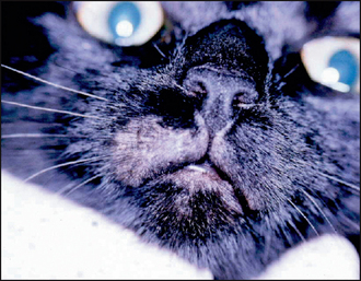
In this case the examination was performed wearing disposable gloves, because from the history dermatophytosis was high on the list of differential diagnoses. A dermatological examination in this case revealed a single expanding patch of alopecia with broken hairs at the periphery on the upper lip (Fig. 28.1). The lesion surface was scaly but non-inflammatory. The general physical examination was within normal limits.
CASE WORK-UP
A diagnosis is made on the history, clinical signs and laboratory tests. In-house screening tests may be carried out to demonstrate the presence of a dermatophyte but the services of a commercial laboratory are likely to be required to speciate the fungus. Knowing the species will help in formulating a strategy both for treatment and control of the fungus within the environment. In-house screening tests include Wood’s lamp examination (Fig. 28.2), direct microscopy and culture on dermatophyte test medium (DTM), and are discussed in detail in Chapter 2. All these tests are dependent on good technique for their success and have their pitfalls.
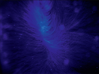
Figure 28.2 Example of apple green fluorescence of M. canis-infected hairs on Wood’s lamp examination.
The following tests were performed:
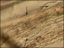
Figure 28.3 Microscopic examination of hair plucks in liquid paraffin mount showing ectothrix arthrospores (arrowed).
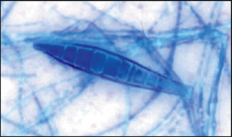
Definitive diagnosis is made on the cultural characteristics and on the microscopic morphology of the fungal hyphae and macroconidia. Microsporum canis appears as cotton wool-like white colonies and the reverse side of the growth medium has a yellow–orange pigment that turns brown. The morphology of the macroconidia is determined by the flag technique. Take a small square of clear adhesive tape and place it on the surface of the colony using a pair of rat tooth forceps. Then place the tape on a microscope slide on which there is a drop of lactophenol cotton blue stain (do not forget to disinfect the instrument with heat or chemical disinfection). Microsporum canis macroconidia contain six or more cells and are spindle shaped with thick walls which form a knob at one end (Fig. 28.4). This technique can also be used on DTM medium to look for macroconidia.
Stay updated, free articles. Join our Telegram channel

Full access? Get Clinical Tree


