11 Dental Radiology
When you have completed this chapter, you will be able to:
• List and describe the indications and contraindications for performing intraoral radiography.
• Differentiate between AC and DC dental radiographic units and describe criteria for choosing a dental radiographic unit.
• Describe the structure of intraoral radiographic film.
• List and describe the components of digital veterinary dental radiology systems.
• Differentiate between parallel and bisecting-angle dental radiographic techniques and describe when each would be used.
• List and describe the views needed for a complete intraoral radiographic study.
• Describe common complications in dental radiology related to improper exposure, positioning, and developing.
• Describe the technique for chairside processing of dental radiographic film.
• Describe normal radiographic anatomy of dogs and cats and common abnormalities that may be seen with radiographic evaluation.
Indications for Dental Radiography
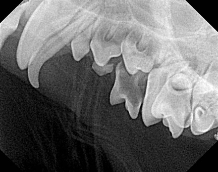
Unerupted or Impacted Teeth
In young patients, radiographs help the practitioner determine whether unerupted or impacted teeth are present (Figure 11-1). An impacted tooth is an unerupted or partially erupted tooth that is prevented from erupting further by any structure. Dental radiographs are taken in all patients to evaluate the status of root and tooth when the tooth is missing or partly erupted. The practitioner may discover that the patient is edentulous or has a dilacerated or an embedded tooth. Edentulous is the absence of teeth. A dilaceration is an abnormally shaped root resulting from trauma during tooth development (see Figure 2-16). An embedded tooth is a tooth that is usually covered in bone, has not erupted into the oral cavity, and is not likely to erupt.
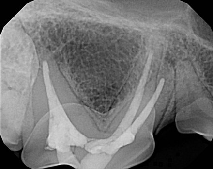
Endodontics
Radiographs allow the practitioner to evaluate the effectiveness of endodontic therapy and to study radicular health and size before, during, and after endodontic therapy (Figure 11-2). Chapter 13 has illustrations of endodontic disease before and after treatment.
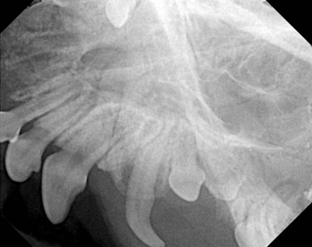
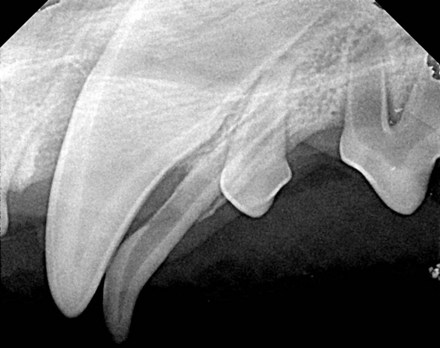
Exodontics
Dental radiographs are indicated before extractions for diagnosis and evaluation of possible complications. Dental radiographs are obtained during the procedure to determine the presence of retained roots and other complications. Radiographs are indicated after extraction to ensure completeness of the procedure. Figure 11-3 shows a preoperative radiograph of a persistent deciduous tooth. The entire root must be extracted. Figure 11-4 shows multiple retained deciduous teeth. The canine tooth will require elevation to extract. The second, third, and fourth premolar roots have resorbed and are being held in place by attached gingiva. They should be removed for the patient’s comfort.
Dental Radiographic Equipment and Materials
Dental Machines
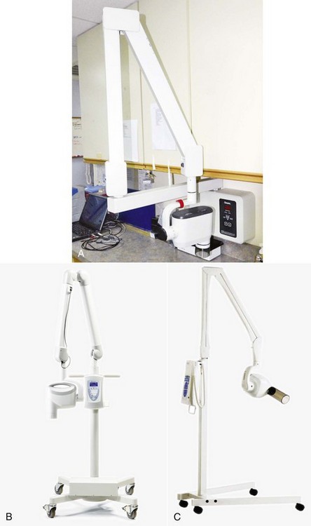
Dental x-ray units may be mounted on the wall (Figure 11-5, A), on stands (Figure 11-5, B and C), or handheld (Figure 11-5, D). The advantage of a wall-mounted unit is that it has a smaller footprint than stand-mounted units. However, wall-mounted units cannot be moved from room to room. Stand-mounted units take space in the dental operatory but can be moved around to different rooms or tables. Handheld units tend to be less powerful than stand-mounted (floor-mounted) units.
Intraoral Film
Description
Most intraoral film comprises a series of layers. A plastic coating covers the external portion. Between the plastic coating and the radiographic film is a layer of paper. The next layer is the radiographic film, sandwiched by another layer of paper. Finally, a layer of lead is followed by another layer of paper that can be peeled from the plastic coating. Some manufacturers have combined the lead and paper layers (Figure 11-6).
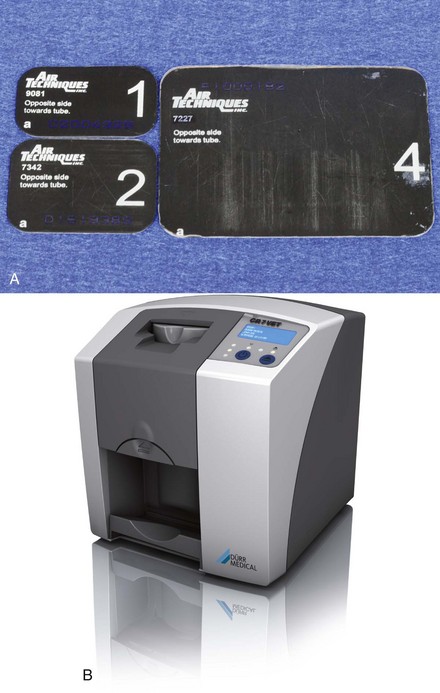
Digital Radiology
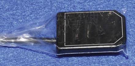
One digital x-ray system, which is known as direct or digital radiology (DR), uses a sensor (Figure 11-7) to capture the image. DR sensors have almost unlimited uses, unless one does bad things to them such as drop them, soak them, or allow animals to bite them. Another digital system has a phosphorous plate (Figure 11-8, A) and processing unit (Figure 11-8, B); this system is known as indirect or computerized radiology (CR). In either case, a laptop or desktop computer is used to process and read the image on a computer screen. While there are differences in appearance, the function is very similar for either of these systems. One of the most important components to the whole system is the computer that is being used to read the image. Both the graphics card and monitor must be high quality and capable of image reproduction.
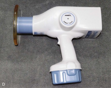
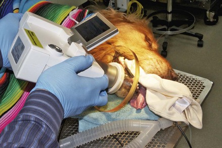
A unique dental unit is a combination handheld unit, digital sensor, and viewer (Figure 11-9).
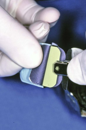
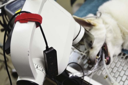
The equipment also must be protected because they are very fragile and subject to damage from being dropped or bit or from water getting inside them. Protective covers should be placed on the unit and secured (Figure 11-10). Another trick is to use Velcro to attach a wire from the sensor to the x-ray arm (Figure 11-11).
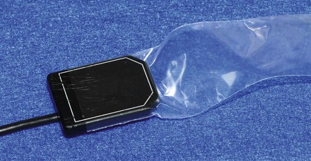
The protective cover will not protect the sensor from animal bites. The patient must be completely anesthetized. Even the slightest flexing may damage a sensor (Figure 11-12).

When it comes to actual application, a live patient is not the place to practice taking radiographs. Skulls (see www.skullsunlimited.com) should be used to perfect the technique. Although children’s play dough is not used in clinical practice, it can be used to position the skull and sensor. In practice, a clean washcloth, laparotomy pad, or gauze can be used to position the sensor.
Stay updated, free articles. Join our Telegram channel

Full access? Get Clinical Tree


