CHAPTER 74 Dental Nerve Blocks
Dental nerve blocks are useful tools that the practitioner can use to aid in the performance of some of the more painful procedures encountered in everyday equine practice. Extractions of cheek teeth, incisors, and canines; treatment of periodontal disease; surgical removal of mandibular periostitis; and wolf tooth removal are some of the common procedures that can be made less painful for the horse through the use of these simple nerve blocks. Less pain for the horse translates into an easier job for the practitioner and less sedation for the horse. The type of local anesthetic used is a matter of personal preference; lidocaine HCl 2% and mepivacaine HCl 2% have fast onset of action and duration of action of 90 to 200 minutes and 120 to 240 minutes, respectively. Proper preparation of the area and use of sterile technique are essential.
TRIGEMINAL NERVE
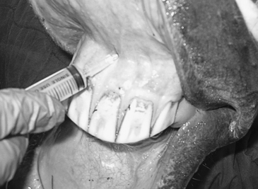
The trigeminal nerve provides the main sensory innervation to most dental structures via two of its three major branches, the maxillary nerve and the mandibular nerve. The maxillary branch of the trigeminal nerve branches into the greater and lesser palatine nerves before entering the maxillary foramen. These pass through the greater palatine canal and the sphenopalatine foramen to innervate the mucosa and glands of the hard and soft palate, respectively. Just after entering the maxillary foramen, the maxillary nerve sends off branches ventrally to innervate the maxillary cheek teeth. It innervates the mucosa of the paranasal sinus and branches before leaving the infraorbital canal to innervate the upper incisors and associated mucosa via branches that continue through the incisive bone and emerge through the incisive canal. The incisive canal is palpable at the junction of the gingiva and labial tissue, dorsal and central to the central upper incisors (Triadan system 101,102). The small maxillary nerve branches that exit here can be desensitized by deposition of 2 to 3 mL of mepivacaine just under the mucosa over the incisive canal (Figure 74-1). This block may be used in conjunction with the infraorbital block when painful procedures involving the incisors are to be undertaken and enable more complete desensitization of the area.
MAXILLARY NERVE
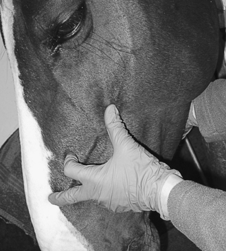
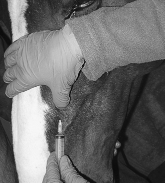
The maxillary nerve leaves the infraorbital foramen as the infraorbital nerve and can be desensitized at that location to provide local anesthesia to the incisors, canines, maxillary bars, and maxillary cheek teeth. The foramen is located halfway along a line from the rostral point of the facial crest to the nasoincisive notch (Figure 74-2). The nerve can be palpated by reflecting the bandlike levator nasolabialis muscle dorsally. The opening of the foramen can be palpated as a shelf, and the nerve can be palpated as it leaves the foramen. A 1½-in. 22-gauge needle is introduced just rostral to the foramen, and a bleb of mepivacaine is deposited as the needle is advanced into the foramen (Figure 74-3). The horse should be heavily sedated or anesthetized and an assistant should prevent the horse’s head from jerking suddenly upward as the needle inevitably comes in contact with the large, broad nerve. Unsedated or lightly sedated horses will react violently to this block. Care must be taken to avoid injury from rearing or striking. Mepivacaine (5 to 10 mL) is deposited in the canal as the needle is being moved retrograde while the opening of the foreman is occluded with the thumb. In cadaver trials, 10 mL of methylene blue traveled the entire length of the infraorbital canal to the maxillary foramen when injected while the foramen was occluded with the thumb. With this technique, complete anesthesia of the upper arcade can be achieved and a block at the maxillary foramen ventral to the orbit becomes unnecessary.
Approaches to the maxillary nerve as it enters the maxillary foramen include a dorsal approach from the caudal aspect of the most dorsal portion of the zygomatic arch (Figure 74-4). A 3½-in. 20-gauge spinal needle is directed medial to the globe and in a slightly rostral direction and advanced to the hub. Two other approaches make use of sites dorsal and ventral to the zygomatic process of the temporal bone (Figures 74-5 and 74-6
Stay updated, free articles. Join our Telegram channel

Full access? Get Clinical Tree


