Cytology of Inflammatory or Neoplastic Masses

16 Cytology of Inflammatory or Neoplastic Masses


Role of Cytology in Diagnosis


Cytologic evaluation of most lesions has become a very common part of diagnosis. Minimal equipment is needed, making cytology possible in any practice. A nearly immediate cytologic conclusion eliminates waiting one or more days for a histologic diagnosis. Cytologic diagnosis gives a cellular or morphologic interpretation to abnormal structures identified by ultrasound or other diagnostic imaging. Fine-needle aspirates are minimally invasive and minimally stressful for patients. Cytology may save the expense of anesthesia and surgery. This chapter discusses cytologic evaluation of masses. Cytologic analysis of fluid, vaginal secretions, semen, urine sediment, respiratory tract material, and specific organs is described in other appropriate chapters.


Cytology can alter or eliminate surgery. Obviously benign masses, such as lipomas and epidermal inclusion cysts, need not be removed immediately. When they are removed, a close resection (i.e., “shelling it out”) is adequate, whereas wide excision is indicated for potentially malignant neoplasms. Any cytologic evidence of malignancy (e.g., possible carcinoma or sarcoma) dictates a search for metastasis before surgical removal of the mass. Evidence of metastasis is a contraindication for surgical removal. Diagnosis of a systemic neoplasm such as lymphoma usually indicates chemotherapy rather than surgery. Neoplasms that are too vascular or invasive for surgery may require cytology for diagnosis instead of histopathology. If a specific tumor diagnosis is made, specific treatment may be started early. Cytologic diagnosis should be considered a tentative diagnosis of neoplasms and histologic diagnosis should be used for more specific classification of the type of neoplasm and degree of malignancy. Some lesions, such as granulomas and granulation tissue, often look malignant on cytology.


Cytology has limitations. Histopathology is usually more diagnostic and definitive, because more information (i.e., tissue architecture) is available from a histologic section than from a variable number of cells on a cytologic smear. Malignancy of certain types of neoplasms (e.g., canine mammary tumor, perianal gland tumor) is best identified by histologic evidence of invasion of tumor cells into normal tissue, and this is seldom documented with cytology. Although cytology gives a definitive diagnosis in many cases (e.g., infectious diseases, mast cell tumor, lymphoma, lipoma), cytology often gives only a general diagnosis in other cases (e.g., epithelial mass with prominent cytologic evidence of malignancy). Most neoplasms require histologic evaluation for a definitive diagnosis and best estimate of malignancy. Radical action (e.g., euthanasia) should await confirmed diagnosis.


Of 147 skin tumors, only 105 (71%) cytologic diagnoses agreed with histologic diagnoses.7 Exceptions exist in which cytology is as diagnostic as (or more diagnostic than) histology (e.g., individual cell detail of leukemias is more diagnostic than tissue patterns). Cytology was correct in 60 of 64 round cell tumors and occasionally more diagnostic than histology.6 Cytologic and histologic diagnoses agreed on all of the following tumors: 37 mast cell tumors, 11 melanomas, 2 histiocytomas, and 1 cutaneous lymphoma.7 Accurate cytologic diagnoses were also made for squamous cell carcinomas, lipomas, and metastasis to lymph nodes.


Cytologic Techniques


Sample Collection and Slide Preparation


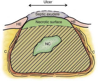
Proper sample collection and slide preparation are absolutely necessary and are a common limiting factor in cytologic diagnosis. Cytologic diagnosis requires an adequate number of cells with good morphology that represent the mass. A fine-needle aspirate (FNA) should be obtained from an area of the mass that reflects the primary problem. The site being sampled determines what material is collected (Figure 16-1). An impression smear from the surface over a mass often has only exudate, bacteria, and necrotic or reactive cells that do not reflect the primary mass. Similarly, a wash over the mass (e.g., nasal flush, bronchoalveolar lavage [BAL]) often collects only exudates and reactive cells on the surface. Pus (septic exudate) may be misleading and suggest the mass is an abscess. FNA samples of deeper tissue are more likely to be diagnostic.


Epithelial cells at the edge of ulcers appear anaplastic, because they are actively proliferating to cover the ulcer (e.g., corneal ulcers can cytologically resemble a squamous cell carcinoma). Ulcerated surfaces often have secondary inflammatory and septic changes. Malignant-appearing cells from deep under a surface are much more likely to truly indicate that the mass is malignant. Soft centers of a mass may be necrotic or hemorrhagic, so a smaller firm mass or a more viable-appearing area at the edge of a mass may be more diagnostic.


An FNA need obtain only one or a few small drops of fluid to streak out similar to a blood smear. Too much aspiration can cause bleeding and hemodilution of the sample. FNA means using a 22- to 20-gauge needle. Vacuum may be applied to a 5- to 10-ml syringe after the needle has penetrated the mass, and the vacuum should be maintained while one passes the needle tip back and forth through the mass; then the vacuum should be released and the needle withdrawn. Cells in the needle then need to be gently placed on a smear and not shot out of the needle as a spray onto the slide. Small droplets of fluid dry too quickly to streak out into a monolayer. They remain as thick drops with poor cell morphology. The needle should be removed from the syringe and the syringe filled with air. The needle is reapplied, and a drop of fluid is gently expressed on a glass slide. The drop is then quickly drawn out into a smear with a coverslip or another slide. The needle can also be used to draw out the drop into thin monolayer extensions (so-called “starfish design”).


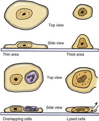
Cells in a thin area (monolayer of cells) spread out flatly and expose a large surface area to view (Figure 16-2). If the smear is thick, cells are supported more upright on the slide and have a smaller diameter. Cells in thick areas are taller (thicker) and thus stain darker, often too dark to see any detail. Protein-rich fluid, necrotic debris, or ultrasound gel surrounding cells interferes with staining. If fluid is viscous, a squash preparation may help to get a thin smear. A drop of fluid is placed between two slides; while the drop spreads to its maximum diameter, the slides are slid apart while the two surfaces are in contact with each other. This creates a smear on each slide. Lymphoid cells and cells from necrotic centers are very fragile and easily lysed beyond recognition. Use of a coverslip for a squash preparation or blood smear–type smear often causes less damage to cells.


A nonaspirate (capillary) technique is easier to perform and yields less bloody samples of equal or greater cellularity, especially from highly vascular tissues (e.g., liver aspirates).9 The technique described here is a modification of the nonaspiration technique used by Dr. Rick Cowell while at Oklahoma State University. Air (i.e., 5 ml) is aspirated into a 10-ml syringe, and a 22-gauge needle is attached. The syringe is held at the base of the needle to allow for better control, and the mass is stabilized with the operator’s free hand. The needle is introduced into the mass and rapidly moved back and forth along the same tract five or six times. Negative pressure (i.e., aspiration) is not applied. The cells are collected by shearing and capillary action. The needle is withdrawn from the mass, and collected cells are quickly but gently expelled onto a clean glass slide by depressing the plunger. The collected material is then spread out as discussed earlier. A common error is the “shotgun spray” in which the small volume of fluid in the needle is sprayed on to the glass as small droplets (like the pattern of shotgun shot hitting a target). These small thick drops dry too quickly to be drawn out into thin smears. Instead, the operator should touch the end of the needle to the glass and gently express the contents out as one drop that is then quickly and gently spread it out as a thin smear. Generally, only one smear can be made from each collection attempt. Therefore three or four collections from different sites should be taken. An alternative is a “packing” technique where only a needle (without syringe attached) is passed through a mass to pack the needle with cells with no aspiration.


If part of a mass is surgically removed, impression smears of representative areas of the mass for cytology should be made before placing the biopsy in formalin. One should use a freshly cut surface and blot off excessive blood and protein-rich fluids. The operator should touch the surface to a glass slide without twisting or rubbing motion. But if no cells are found on those smears or the mass feels hard, then cells should be aggressively scraped off from the surface with a blade and those cells streaked onto a slide. Fibrous masses usually do not release cells easily and may need to be scraped with a scalpel to obtain enough cells for diagnosis. The moist material on the blade is streaked on glass slides.


Patient identification and other information must be noted on the slide. An adequate description is needed for a cytologist, who has not seen the animal or lesion, to interpret the results and provide a useful answer to the veterinarian submitting the sample. The description should include the site and a description of the lesion (e.g., “packing technique from an enlarged liver” or “FNA of a skin tumor near the anus”). Such description is preferable to a common request of “What kind of cells are present?” without describing what lesion in what part of the animal was sampled. A specific question allows a specific answer. If one or more sites were sampled, one should note how each slide relates to the sites sampled. Dates are needed if samples were taken at different times. Slides should be labeled with lead pencil on a frosted end–type slide. Printer-generated paper labels for marking test tubes cause problems on cytology slides. When the slide is stained, the paper label may also be so darkly stained that no word is legible. Computer labels are too large for the slide to fit on the microscope stage and have glue on them. When the excessive paper is removed, glue remains so the slides stick to the microscope stage, preventing that slide or subsequent slides from being movable. Thin two-slide cardboard boxes mailed to a cytologist often arrive with crushed glass slides, because the envelope is machine cancelled even when they are marked “Hand Cancel.” Therefore, slide containers that are too large (e.g., rectangular plastic box) to fit through the post office’s automatic stamp canceling machine should be used for mailing.


Smears are routinely air-dried for Wright-type stains and new methylene blue (NMB) staining. Alcohol fixation is required for Papanicolaou’s (Pap’s) or Sano’s stain, which few cytologists use. Air-drying smears slowly in a moist environment may cause cell distortion. A hair dryer may be used to speed drying if the problem persists. The dryer should be held far enough away from the smears to avoid “cooking” cells. There is no need to “flame” smears for cytology of neoplasia, which requires optimal morphology. However, flaming slides from waxy ear swabs or colonies from blood agar plates for Gram staining is helpful. Smears should not be stored in refrigerators or with exposure to dust, molds, pollen, and flies. Flies eat unstained cells. One should handle slides only on the sides of the glass, because squamous cells from fingerprints can contaminate smears and interfere with interpretation.


Smears exposed to formalin often have excessive blue staining with Wright stain. Tissue samples used for impression smears should not have been placed in formalin before the slides were made. Formalin should not be stored near the stains or smears. Formalin should not be submitted in the same package with cytologic smears, where the fumes may act on unstained cells. Heparin anticoagulant also causes a blue discoloration of Wright-stained cells.


Stains


A modified Wright-type stain and NMB are usually adequate for diagnosis. The “quick” stains used today may be called “Wright stains” but are really not Wright or Giemsa (i.e., true Romanowsky’s) stains. Quick stains such as Diff-Quik and Hemacolor have blue and red dyes to give staining characteristics similar to classic Romanowsky’s stains. The blue dyes stain acidic structures such as nucleic acids (deoxyribonucleic acid [DNA] and ribonucleic acid [RNA]) in nuclei blue to purple. Red dyes stain basic structures such as proteins (e.g., hemoglobin in erythrocytes) red to orange. Changes in pH in rinse water or the sample can affect staining characteristics. A quick stain such as Diff-Quik can be adjusted to color cells on the smears more blue or red or darker. The blue and red dyes are in separate jars; by increasing or decreasing the number of times one dips the slide in a color, the intensity of blue or red is increased or decreased. Diff-Quik stains distemper inclusion bodies better on blood smears than some “Wright stains.” Water-based “Wright stains” may fail to stain granules in mast cells and basophils. Ear swab smears should be stained in separate staining jars for only ear swabs because the stains can quickly be contaminated with yeast, which are transferred to smears from other patients.


NMB is a monochrome stain with variably intense blue staining. A “wet mount” is made by placing a drop of NMB on an air-dried smear and applying a coverslip. To prevent retention of an air bubble over part of the smear (usually the most diagnostic area), the coverslip should be used to gently pull the drop of NMB over the cells to moisten them before slowly applying the coverslip. Staining is immediate. NMB stains nuclear material well and demonstrates distinct chromatin patterns (Figure 16-3). This nuclear detail is very useful in evaluating malignant criteria. The transparent nature of NMB is a major advantage, because one can see through thicker tissue fragments that would be too darkly stained with a Wright stain. The microscope can be focused through different depths of tissue fragments to judge individual cell detail and architectural patterns in three dimensions. Tissue fragments are often the most diagnostic material on smears from neoplasms but stain too darkly to evaluate with a Wright stain. The fragments are like tiny biopsy sections that allow evaluation of how cells were oriented in the mass. These architectural patterns help identify the tissue type. One can evaluate adjacent cells for true variability suggesting malignancy, compared with the variability of isolated cells on a smear that may have come from different areas or cell types in the mass.


In summary, NMB is excellent for nuclear detail, thick tissue fragments, and most fungi. Wright stain is excellent for inflammatory lesions, because the stained appearance of leukocytes (white blood cells [WBCs]) is similar to that in blood smears and bacteria consistently have a characteristic dull blue color (Figure 16-4). Although a Wright stain is not as good as NMB for nuclear detail, it is acceptable for evaluating tissue cells for criteria of malignancy and excellent for bacteria and most fungi (Figure 16-5). Use of both Wright and NMB stains for the same lesion works well, because different characteristics of the cells are illustrated by different stains.


Other stains may be used. Sudan stain is useful for diagnosis of fatty liver, chylothorax (Figure 16-6), aspiration pneumonia (Figure 16-7), or lipid granulomas. A drop of Sudan stain (or other neutral fat stain) is drawn over a smear to stain the cells and background selectively for lipid. Excess stain is poured off and then the smear is counter-stained with a drop of NMB to show adequate cell detail. NMB stains the nucleus and other cell structures blue, and the Sudan stains neutral fat red.


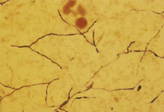
Veterinarians sometimes request that their cytologic samples are Gram stained because different antibiotics are used for gram-positive versus gram-negative bacteria. However, Gram staining is absolutely not recommended for cytologic smears. Gram staining is inconsistent for bacteria in exudate. In thick smears the bacteria may not decolorize, creating a false impression that the bacteria are gram positive. In thin smears, bacteria may decolorize too much, suggesting they are gram negative. Gram staining is not sensitive for screening cytologic smears for gram-negative (i.e., red) bacteria in low numbers in a red, proteinaceous background (Figure 16-8). Wright-Giemsa–type stains consistently stain bacteria blue and are greatly preferred for finding bacteria on cytologic smears. Wright-Giemsa stains allow easier detection and definition of size and shape. In general, coccoid bacteria are likely gram-positive Staphylococcus and Streptococcus. Gram staining is best restricted to bacteriology laboratories where smears are consistent in thickness and staining is interpreted daily on bacteria where the final classification is determined daily. Gram stain differentiates gram-negative from gram-positive bacteria well on uniformly thin smears from cultures on blood agar plates. Acid-fast stain is rarely needed, because mycobacterial infections are uncommon (see Figure 16-8). Mycobacteria are unique in that they do not stain at all with Wright-Giemsa stains because of their waxy coat (Figure 16-9).


Pap’s stain is often used in human medicine (cervical swabs) and rarely in veterinary practice. Pap’s stain has advantages; however, this author usually was able to make a diagnosis with NMB and Wright-stained smears and write out the report before the technologists could finish staining smears with Pap’s stain. Therefore we ceased using Pap’s staining. Specific malignant criteria have been established for cells stained with these stains. Pap’s stain is a transparent stain that permits evaluation of thick tissue fragments and fine evaluation of nuclear characteristics. Smears need to be immediately fixed in alcohol before Pap’s staining.


Microscopes


A good-quality, well-maintained microscope is needed for cytology. An ergonomic binocular microscope is more comfortable for long viewing periods. Four objectives are recommended: a 4× and a 10× objective are used for scanning a smear and quickly finding likely diagnostic areas, which are then examined with 50× oil and 100× oil objectives for fine details. A properly equipped, good-quality microscope is expensive but will have multiple uses in a clinic and should last a lifetime. The microscope cost per each slide is examined below.


A 50× oil plan achromat objective (i.e., 40× to 60× oil) pays for itself ($350 to $1000) by the time it saves to examine a slide. Magnification is sufficient for most detail, and more cells can be seen in a shorter time. “Plan” means the whole field of view is in focus. Having more cells in the larger field of view allows better comparison of variations among cells and easier identification. Achromat corrects spherical aberration for 1 color. Apochromat corrects for 3 to 4 colors but is more expensive and not needed for diagnosis. A 50× oil objective avoids the need to coverslip smears.


Wright-stained cells observed with most 40× high-dry objectives appear fuzzy, because the cells are surrounded by air. Using mounting media with a coverslip or oil eliminates the air-cell interface and allows good cellular detail with a high-dry lens. Adding a drop of oil is much faster than permanently coverslipping smears. Oily smears are messy to store and less permanent, however. High-dry 45× objectives are easily contaminated with oil on smears and the convex lens is difficult to clear of oil. A microscope with only oil objectives avoids this problem and loss of time.


Oil should be removed from lenses at the end of a work period, because oil can penetrate behind some lenses to render them useless or dry to become a hard coating on the lens. Immersion oil is not removed by alcohol but is removed by gasoline or xylene, which should be used instead of alcohol for routine cleaning. Kimwipes are lint-free tissues that are satisfactory for cleaning microscope objectives not used for photomicroscopy. Kimwipes absorb oil better than does lens paper and thus clean more effectively. Concave lenses require a cotton-tipped swab moistened with gasoline to clean the recessed area. Final polishing of the objective lens should be with lens paper. One should clean filters, light sources, stage, and condenser as needed. Complete covering of the microscope prevents accumulation of dust in places hard to clean. Sharp vibrations (e.g., dragging the microscope along the surface of a desk, setting it down hard) should be avoided. This can knock the prism out of alignment and cause a double image.


The condenser must be in the proper position for optimal detail (e.g., for finding small bacteria). The condenser is near the optimal setting (i.e., Köhler illumination), if it is close beneath the glass slide on the stage. More specifically, several simple steps should be performed daily. While at a high magnification (e.g., 40× objective), one should focus on the cells on a smear. The field diaphragm at the bottom of the microscope is completely closed, and the condenser is moved slightly up or down until the circle of light in the field has sharp edges. The condenser should be then left at this optimal height. One should then center the circle of light to the center of the field and reopen the field diaphragm fully so that no shadow appears in the field. The diaphragm in the condenser can be adjusted to optimize contrast but, more simply, it should be fully open for most users. High-quality oil objectives available today, function well with the condenser diaphragm fully open. A common error is closing the aperture diaphragm in the condenser too much, causing too much contrast. This is usually done (incorrectly) to reduce light intensity. The proper way to reduce light intensity is to use a neutral-density filter, a dimmer bulb, or a more controllable light source rather than to cause poor cell detail. Improved contrast may be obtained by slightly closing the aperture diaphragm in the condenser, but this should be left to experienced microscopists using photomicroscopy.


When morphologic detail is not needed and the goal is only to easily find objects such as parasite ova in a fecal exam, urinary casts in urine sediment, or platelets in a hemocytometer, the condenser is moved to the lowest position that still provides adequate illumination. This position gives clear structures more contrast and they are easier to find.


Some common problems with microscopy include placing the slide upside down on the stage. In this case one can focus on cells at medium magnification (i.e., 10×, 40×) but not with the 100× oil objective, because cells are on the underside of the slide. If the coverslip or mounting medium is too thick, one also cannot focus at high magnifications. If the fine focus will not turn any farther in the direction needed, one should adjust the coarse adjustment past the plane of focus needed, and then turn the fine focus knob in the opposite direction to regain focusing ability. If the cells look refractile and have poor detail, the lighting is probably wrong. In this case one should adjust the microscope to Köhler illumination. If cells are in focus with the 100× objective but not the high-dry 45× objective, the 45× objective is likely contaminated with oil and must be cleaned.


Cytologic Conclusions


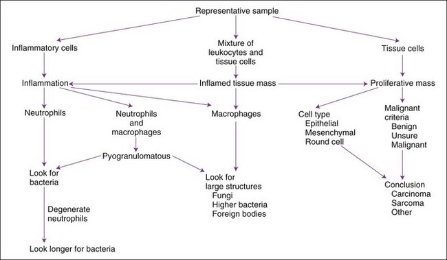
The usual composition of a mass is a proliferation of tissue cells, an accumulation of inflammatory cells, or both. Miscellaneous masses include hematomas, cysts, or focal areas of necrosis. A general approach to cytologic interpretation of a case is simplified in Figure 16-10. More complete description of many cytologic diagnoses is available.11


Slide Reading Approach


Most American veterinarians have ample microscopy, histology, and pathology training, so most can learn to make many cytologic diagnoses as long as they recognize their limitations and continue to learn from their cases. Cytologic evaluation may be performed on excised masses and then the descriptions and conclusions may be compared with histopathology reports. Cytologic evaluation is a visual task, so one should obtain one or more cytologic atlases for frequent reference in the lab.2,5,11 An organized approach to an aspirate or impression smear of an abnormal mass is necessary for consistent conclusions. A summary of steps follows, and details are provided in later discussions.


One should first determine if the cytologic specimen likely represents the lesion. Adjacent structures may be sampled inadvertently. For example, a common error is to aspirate the salivary gland instead of the submandibular lymph node. The salivary gland has normal, mature acinar and ductal structures with foamy epithelial cells. The conclusion should be that the sample was not representative, not that an adenoma or metastatic carcinoma was present. Other examples include inadvertently sampling the liver while obtaining “thoracic” aspirates and having the needle pass all the way through a small mass and to only aspirate subcutaneous (SC) fat. One may contaminate a cystocentesis urine sample with gut bacteria by inadvertently puncturing the intestine. Correct conclusions often require intuition and experience.


The initial effort should be to screen smears grossly for those areas most likely to be diagnostic. Smears that likely have small tissue fragments appear granular and should be stained with NMB or other semi-transparent stain. Smears that stain dark blue are the most cell-rich smears and are most likely diagnostic. The intense blue color is because they have the most nuclei. Hemodiluted smears with few nucleated cells appear orange (like a blood smear) with Wright’s stain, suggesting reduced chance of diagnosis.


Too often one goes too quickly to the 100× oil objective and stays at that power until fatigued. The scanning objectives (4×, 10×) must be used first, and often, to locate productive areas of the smear, which are then evaluated with an oil objective power (50×, 100×). Promising areas are thin and have intact, well-stained individualized cells. Cells poorly stained with Wright stain have an altered, pale, diffuse, blue color. The color and streaked-out appearance of necrotic, lysed cells indicates an area to avoid. Diagnostic structures (e.g., bacterial and fungal colonies) may be rare and isolated, so one should invest one’s time to scan smears and not waste time at high magnification in a few areas. Similarly, tissue fragments that have the valuable architectural patterns are irregularly distributed and found by scanning. Tissue particles appear as dark granules often at the feathered edge.


An accurate and complete description makes conclusions easier. Performing a differential count of cells with a hematology differential counter forces the cytologist to classify each and every cell and not be biased by prominent cells such as eosinophils, plasma cells, and large cells that seem more numerous than small cells such as lymphocytes. One should not expect to identify all cells. It is common to have a few unidentified cells (i.e., fibroblasts, monoblasts, lymphoblasts) in inflammatory masses, and these few cells may look immature and have cytologic characteristics of malignancy. If they are few, they may be accepted as reactive cells secondary to inflammation. Recall that a confident cytologic report of a malignant neoplasm often leads the veterinarian to kill the patient without further testing that may give a better diagnosis. No cytologist likes killing dogs and cats.


Inflammatory Masses


Inflammation is diagnosed much more frequently and easier with cytology than is neoplasia. Cytologic diagnosis simply requires an adequate number of inflammatory cells. The number of cells sufficient for diagnosis varies with the sample. A rare plasma cell and phagocytic macrophage aspirated from inside the eye indicates inflammation, whereas thousands of neutrophils are found in pus. In hemodiluted samples, one considers the number and type of WBC usually found in blood. Blood has about a 500 : 1 ratio of red blood cells (RBCs) to WBCs, with mainly neutrophils and lymphocytes. More WBCs (e.g., 20 : 1 or 1 : 20 ratio) or the presence of a WBC not found in blood (e.g., plasma cells, phagocytic macrophages) is used with hemodiluted samples to diagnose inflammation (e.g., hepatic cytology). Based on predominant WBC type, different terms are used and different causes are suspected.


Neutrophilic Inflammation


Neutrophilic infiltrates (e.g., exudation, suppuration, abscess formation, purulent inflammation) are so frequently seen that they are almost synonymous with inflammation. Neutrophils are the most motile WBCs and the first to infiltrate an area or exude out on a surface. Some call neutrophilic inflammation “acute inflammation,” even though neutrophils may be prominent in chronic but active inflammation. Therefore the term acute may refer to a cell type (e.g., predominance of neutrophils) and not always to a time interval. Pus is proteinaceous fluid with many neutrophils and cell debris. Aspiration readily collects this fluid material; therefore many neutrophils are often present on smears. Other cells in an inflammatory mass may not exfoliate as easily (especially fibroblasts) if scarring and fibrosis are present. Neutrophils are associated with bacterial infections and some yeast infections (e.g., Candida), but nonseptic causes include immune-mediated processes (e.g., lupus polyarthritis) and chemical irritation (e.g., pancreatitis, bile peritonitis). A neutrophil migrating between stratified squamous epithelial cells may indent into the surface of a squamous cell and appear as if it is within the squamous cell when it exfoliates (see Figure 16-2).


Bacterial Sepsis


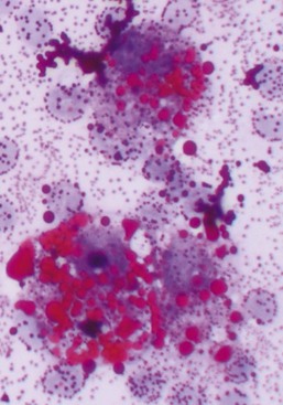
Neutrophilic inflammation indicates a search for bacteria. The best place to search is in cytoplasm of neutrophils (see Figure 16-4). The neutrophil’s cytoplasm is usually clear and free of granular debris. Macrophages often contain phagocytized cell debris that can mimic bacteria. Bacteria are more prominent in the clear neutrophilic cytoplasm, and the phagocytic vacuole may help outline the organism. Bacteria have uniform shapes and sizes, in contrast with granular debris. Formation of uniform pairs, tetrads, and chains identifies structures as bacteria. Rods are more confidently identified as bacteria than cocci. Wright stain precipitate is coccoid in appearance and may mimic coccoid bacteria. However, the irregular size of the precipitate, a more purple color, and a refractile appearance will differentiate stain precipitate from bacteria. Bacteria have a more dull blue color (see Figure 16-4). Stain precipitate may be on neutrophils, suggesting phagocytosis, but will also be elsewhere on the glass slide including where no sample was applied.


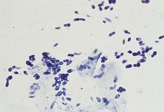
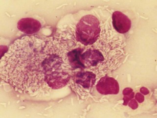
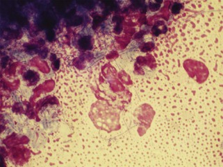
The description of bacteria should include number, location (e.g., free in the smear, phagocytized by neutrophils, or on epithelial cells), appearance, and whether a pure or mixed population is present. These observations permit certain conclusions. For example, a pure population of small cocci in chains within neutrophils from an abscessed lymph node suggests an infection (e.g., Streptococcus), whereas a mixed population of variably sized, large rods and even cocci in neutrophils in abdominal fluid suggests a ruptured gut. Beaded filamentous organisms indicate higher bacteria (e.g., Actinomyces) (Figure 16-11; see also Figure 16-8).


Phagocytosis of bacteria by neutrophils is a better indication that there was bacterial infection than are free bacteria. But even free bacteria combined with neutrophilic inflammation support probable infection, especially if in a fresh sample. Bacteria or fungi free in the background may have been bacterial or fungal contamination in the stain or with older samples. Contamination is especially likely in samples that were moist (e.g., tracheal wash) and stored for hours or days before a smear was made. Bacteria or fungi in a sample without an inflammatory response are usually contamination, with some exceptions (e.g., ear swabs, diabetes, Cushing disease).


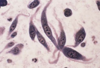
Bacteria on stratified squamous epithelial cells are usually normal flora from a body surface. A normal flora of the oropharyngeal area of dogs is Simonsiella. Finding this characteristic, huge, flat multicellular form (called trichrome) of 12 to 20 bacterial cells indicates at least part of the sample came from the mouth or pharynx. Finding Simonsiella and a mixture of other bacteria on squames indicates that one cannot trust finding bacteria in another area of a transtracheal wash or BAL as indication of infection of the lower respiratory tract. Bordatella bronchiseptica has a predilection for cilia of respiratory epithelial cells, so finding small rods on cilia suggests Bordatella infection (Figure 16-12).


Degenerative Neutrophils


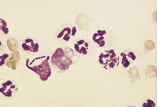
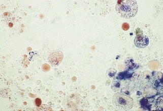
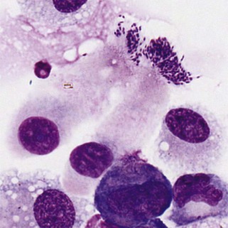
How long should one search for bacteria in a sample with neutrophilic inflammation? Bacteria may be in low concentrations in chronic infections, with antibiotic therapy, and in some samples such as joint fluid and cerebrospinal fluid (CSF). Five to 10 minutes is a reasonable time limit unless something suggests searching longer. For example, one should search for bacteria longer than usual if neutrophils appear degenerate. Bacterial toxins often cause rapid neutrophil death (karyolysis). Degenerative changes in neutrophils suggest but do not prove sepsis. Some bacteria seem less toxic to neutrophils, however, and bacteria may be found in nondegenerate neutrophils (see Figure 16-4).


Morphologically degenerate neutrophils are characterized by swelling of the nucleus (karyolysis) and cytoplasm. Karyolysis appears as a wider, more irregularly shaped, lighter-staining nucleus lacking the dark, distinctly granular chromatin pattern and thin lobulated shape of viable nuclei (see Figure 16-11). Severely degenerate neutrophils may barely resemble neutrophils as they swell and lyse into “globs” of nuclear debris. Degenerative changes caused by bacteria must be differentiated from swelling due to sample storage, trauma to fragile cells during streaking of the smear, or nonbacterial toxic effects (e.g., urine). Inexperienced cytologists tend to over-identify degenerative neutrophil changes by examining damaged cells (see Figure 16-4). One should evaluate only intact, undamaged cells. If the neutrophils with intact cell boundaries appear nondegenerate, lysed neutrophils on the slide are probably artifactually broken rather than degenerate from bacterial toxins.


Nondegenerate neutrophils resemble normal neutrophils in fresh blood smears (i.e., clear cytoplasm; a dark, thin, lobulated nucleus). A lack of bacterial toxins permits cells to live longer. Old neutrophils become hypersegmented and are evidence of a nontoxic environment. Nondegenerate neutrophils die slowly of old age and develop pyknotic or karyorrhectic nuclei. Pyknosis (one very dark, homogeneous nucleus) and karyorrhexis (variably sized dark, homogeneous, round fragments of nuclear material) are evidence for nondegenerate neutrophils.

Stay updated, free articles. Join our Telegram channel

Sep 10, 2016 | Posted by in SMALL ANIMAL | Comments Off on Cytology of Inflammatory or Neoplastic Masses

Full access? Get Clinical Tree

Get Clinical Tree app for offline access