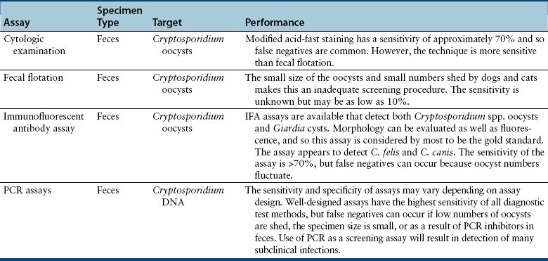
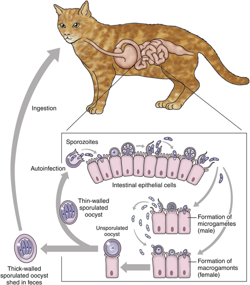
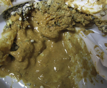
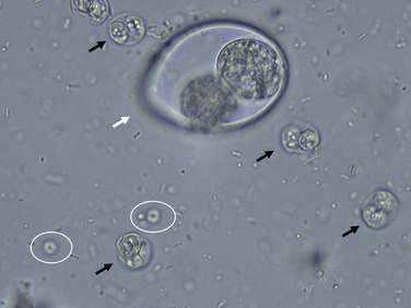
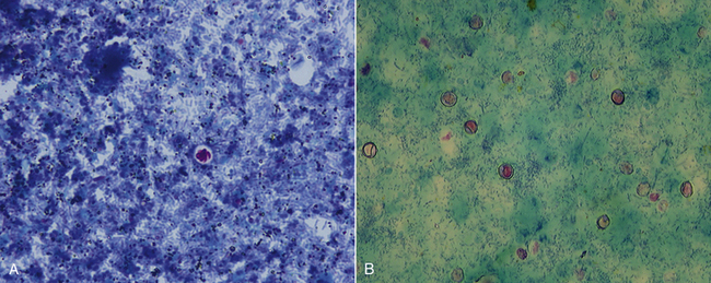
Chapter 81 Cryptosporidium spp. are coccidians that reside in the small intestines and are occasionally associated with disease in some infected hosts. There are now 16 accepted species of Cryptosporidium, and approximately 50 Cryptosporidium genotypes have been described.4 In the past, most mammalian cryptosporidiosis was attributed to Cryptosporidium parvum. However, molecular studies have demonstrated that cats are usually infected with the host-specific Cryptosporidium felis and dogs are usually infected with Cryptosporidium canis.4–16 Occasionally, dogs or cats are infected with C. parvum.17 A mixed infection with Cryptosporidium muris and C. felis was reported in one cat.18 Cryptosporidium hominis is the most common human host–adapted strain and has not been associated with infection in dogs and cats. Cryptosporidium spp. infections of dogs and cats can be quite common, with prevalence rates generally being 2% to 12% in dogs or cats with or without diarrhea, depending on the method of diagnostic testing.19–30 In one study of specimens collected from around the United States, Cryptosporidium spp. DNA was amplified by PCR assay from feces of 29.4% of cats and 15.1% of dogs with diarrhea.24 In a study of cats in shelters in upstate New York, fecal flotation identified oocysts in 3.8% of the cats tested.21 Cryptosporidium felis and C. canis are transmitted among dogs and cats by the ingestion of oocysts in feces from mutual grooming, shared litter boxes, ingestion of contaminated food or water, and possibly ingestion of infected prey species. The oocysts are passed in the feces already sporulated and so are immediately infectious (Figure 81-1). In a study of cats inoculated with C. parvum, oocysts were detected by day 7 and C. parvum DNA was detected by day 2 after inoculation.31 In contrast, C. felis oocysts are first shed in the feces 3 to 6 days after infection. Approximately 20% of the oocysts produced in the intestine are “thin-walled” oocysts that fail to form an oocyst wall. These oocysts rupture within the intestines, and when the sporozoites are released, auto-infection occurs, which allows for rapid amplification of infection. Thick-walled oocysts are passed in the feces, are environmentally resistant, and are the likely source of new infections. Between 1 and 1000 oocysts of pathogenic species such as C. parvum are enough to cause infection in humans.4 FIGURE 81-1 Life cycle of Cryptosporidium spp. Gametogony generates both thin-walled oocysts, which are immediately infective, and thick-walled oocysts, which are shed in the feces already sporulated. Although infection of dogs and cats with Cryptosporidium spp. is common, most infected animals are clinically normal. In prevalence studies, only rarely is there an association between positive Cryptosporidium spp. assay results and the presence of diarrhea.28 Diarrhea is generally more common in young animals.32–46 When diarrhea occurs, it is usually watery, without mucus, blood, melena, or straining, and so is classified as small bowel diarrhea (Figure 81-2). Weight loss may also occur. Co-infection with other protozoans such as Giardia spp. (dogs and cats) or Tritrichomonas foetus (cats) may be associated with more significant illness than infection with just one of the parasites.47,48 The presence of immunosuppressive diseases such as lymphoma, FeLV infection, canine distemper virus infection, or canine parvovirus infection also can potentiate the development of clinical signs of disease. Clinical signs appear to be more likely in cats infected with C. felis than in dogs infected with C. canis. In the cat with a mixed C. muris and C. felis infection, both vomiting and diarrhea were present, and C. muris was identified in the stomach in the presence of inflammation.18 FIGURE 81-2 Characteristic appearance of diarrhea induced by Cryptosporidium felis and Giardia spp. co-infection. The image is from the case example described at the end of this chapter. Little information is available in regard to the pathogenesis of Cryptosporidium spp. in cats and dogs; most information is from what is known in humans and mice after infection by C. parvum.49 After ingestion, Cryptosporidium spp. sporozoites attach to the intestinal epithelium via interactions among multiple parasite ligands and host receptors. The organism resides between the cell membrane and the cell cytoplasm, which may partially explain its resistance to antiprotozoal chemotherapy. The intestinal epithelial cells act as a physical barrier and produce a variety of cytokines and chemokines in response to the pathogen. Mucosal infiltration with neutrophils, macrophages, and lymphocytes in the lamina propria can result. There is upregulation of both proinflammatory and antiinflammatory cytokines, and the ratio of these cytokines likely determines whether diarrhea develops. Cellular immunity mediated by CD4+ and CD8+α/β T cells is an important component for the resolution of C. parvum infection. When it occurs, Cryptosporidium spp. diarrhea is associated with impaired intestinal absorption and enhanced secretion.49 It is possible that susceptibility to cryptosporidiosis in animals could have a genetic basis as suggested for humans.50,51 Specific diagnostic assays for cryptosporidiosis are shown in Table 81-1. Cryptosporidium felis and C. canis oocysts are similar in size; C. felis oocysts are 5 µm by 4.5 µm and C. canis oocysts are 4.95 µm by 4.71 µm (Figure 81-3). However, their small size means that the oocysts are easy to overlook. Modified acid-fast staining of a thin fecal smear can be performed in the small animal practice to aid in the detection of the organisms (Figure 81-4).23,52 Cryptosporidium spp. are generally the only enteric organism of the size just specified that stain pink to red with acid-fast stain. However, in one study, acid-fast staining detected only approximately 70% of Cryptosporidium spp.–infected kittens when a single specimen was tested.52 FIGURE 81-3 Fecal flotation from a cat that was co-infected with Toxoplasma gondii, Isospora felis, and Cryptosporidium felis. The image is magnified approximately 2,000×. The black arrows are sporulated T. gondii oocysts; the white arrow shows Isospora felis; the white circles surround Cryptosporidium felis oocysts. FIGURE 81-4 Acid-fast (Ziehl-Neelsen) stained fecal smears showing Cryptosporidium felis. A, Smear from a cat with diarrhea that shows a single oocyst of C. felis. (From: Marks SL, Willard MD. Diarrhea in kittens. In: August JR, ed. Consultations in Feline Internal Medicine. 5th ed. St. Louis: Saunders; 2006.) B, Multiple oocysts of C. felis that have a more crinkled appearance. The oocysts are approximately 5 µm in diameter and stain pink to red. (From Ettinger SJ, Feldman EC. Textbook of Veterinary Internal Medicine. 7th ed. St. Louis: Saunders; 2011.) Centrifugal fecal flotation is usually performed as part of the initial diagnostic work-up of dogs or cats with small bowel diarrhea.53 Cryptosporidium spp. oocysts are frequently missed because of their small size, and only low numbers of oocysts are passed in infected dog or cat feces (often <500 oocysts/gram of feces). A fluorescein-labeled monoclonal antibody system is available that contains monoclonal antibodies that react with Cryptosporidium spp. oocysts and Giardia spp. cysts (Merifluor IFA, Meridian Biosciences, Cincinnati, OH). In limited studies, this assay appears to detect both Giardia spp. and Cryptosporidium spp. isolates from dogs and cats.12,24,31,52 The IFA provides both a morphologic assessment of the oocysts and immunologic identification, which makes false-positive results unlikely. Fecal ELISA antigen tests for Cryptosporidium spp. are available for use with human feces, but results of these assays have been variable when applied to feces from infected animals.12,52,54 The assays use antibodies against C. parvum in their design, and so the high false-negative rate in animals may reflect antigenic differences among C. parvum, C. felis, and C. canis. PCR assays are currently available to amplify Cryptosporidium spp. DNA from canine or feline feces. These assays appear to be more sensitive than IFA when used with cat feces.24,31,55 Real-time PCR assays are available commercially through some veterinary diagnostic laboratories. Specific PCR assays are available for each host-adapted species, or PCR products generated with genus-specific assays can be sequenced to identify the Cryptosporidium species present. Because Cryptosporidium oocysts or DNA can be detected in apparently healthy dogs and cats, positive test results do not prove a disease association.
Cryptosporidiosis
Etiologic Agent and Epidemiology
Clinical Features
Diagnosis
Microbiologic Testing
Cytologic Examination
Concentration Techniques
Direct Immunofluorescent Antibody Testing
Fecal Antigen Detection by Enzyme-Linked Immunosorbent Assays
Molecular Genetic Techniques
![]()
Stay updated, free articles. Join our Telegram channel

Full access? Get Clinical Tree


Veterian Key
Fastest Veterinary Medicine Insight Engine