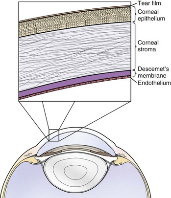
Corneal Ulcers

Chapter 246


Corneal Ulcers


Corneal Anatomy


The cornea and sclera make up the tough and protective fibrous tunic of the eye. The cornea is composed of five layers: precorneal tear film, epithelium with basement membrane, stroma, Descemet’s membrane, and endothelium (Figure 246-1). The cornea can be described as a “fat-water-fat” sandwich; the epithelium and endothelium–Descemet’s membrane are hydrophobic, whereas the collagenous stroma is hydrophilic.


The epithelium of the cornea is protected and nourished by the precorneal tear film, which consists of three layers: (1) a mucus layer produced by the goblet cells of the conjunctiva, (2) an aqueous layer produced by the orbital lacrimal gland and gland of the third eyelid, and (3) a lipid layer produced by the meibomian glands. Functions of the tear film include lubrication of the ocular surface, provision of oxygen and nutrients for the anterior cornea, removal of waste products, immunoprotection (via endogenous components, including immunoglobulin A and lysozyme), and maintenance of optical transparency. A normal, healthy tear film is critical for the maintenance of corneal health.


The corneal epithelium is approximately six to eight cell layers thick, with the deepest layer being comprised of columnar basal cells. The epithelium is nonkeratinized and firmly attached to the underlying stroma by hemidesmosomes. The epithelium has an important function as a physical barrier to prevent the seepage of the precorneal tear film into the cornea. It also acts as a barrier to microorganisms, preserving the integrity of all other ocular structures.


Stroma represents approximately 90% of the corneal thickness. It is composed of well-organized, parallel layers of collagen fibrils, glycosaminoglycans, and a relatively low number of keratocytes. The bulk of the stroma is comprised of water, which makes this corneal layer highly hydrophilic. The precise anatomic arrangement of the collagen fibrils allows light to pass through the cornea without scatter.


Descemet’s membrane is the basement membrane (10 to 15 µm thick) of the corneal endothelium. It is highly hydrophobic and gets thicker with age. The endothelium is a single layer of hexagonal cells lining the inner surface of the cornea. The endothelial cell layer is very active metabolically and plays the major role in maintaining corneal transparency. Adenosine triphosphate–dependent pumps on the endothelial cells help to transport water actively out of the corneal stroma to maintain its relatively dehydrated state.


Physiology of Corneal Transparency


Corneal transparency is maintained by the following five mechanisms:


Loss of corneal transparency results from a disruption of one of these normal physiologic mechanisms. Corneal ulceration results in corneal stromal edema due to loss of the hydrophobic epithelial barrier. Corneal vascularization or pigmentation may develop with chronic corneal ulceration. Keratinization typically is observed at a microscopic level in patients with keratoconjunctivitis sicca (KCS), but it usually is not associated with corneal ulceration. Alterations in the regular arrangement of the stromal lamellae lead to loss of transparency. Scar formation following injury of the stroma results in opacity due to misalignment of the collagen fibers.


Corneal Wound Healing


Wounds that involve the corneal epithelium heal differently than wounds that involve the stroma. Epithelium is mitotically active and quick to repair superficial wounds. Epithelial cells at the edges of the ulcerated area release their attachments to neighboring cells, migrate to cover the wound, undergo mitosis to rebuild the normal epithelial thickness, and then reestablish their connection to the underlying basement membrane. Epithelium migrates at a rate of approximately 1 mm/day from both sides of the ulcer bed. Stromal wounds are much slower to heal. For a wound involving stroma to resolve, epithelium must slide over the defect and keratocytes must be activated into a fibroblast phenotype, migrate to the wound bed, remodel the injured collagen, and synthesize new collagen. This process takes weeks to months to complete. Corneal fibrosis typically results from wounds involving the stroma because newly synthesized collagen does not have the precise anatomic orientation of mature collagen. If epithelialization precedes stromal remodeling, a corneal facet can occur. A facet is an area of thin stroma covered by intact epithelium. It is important to differentiate a healed facet from a descemetocele by carefully observing the fluorescein staining pattern of the lesion. A facet does not retain fluorescein dye, whereas a descemetocele likely shows fluorescein retention in the wall but not in the base of the ulceration.


Diagnosis of Corneal Ulceration


A corneal erosion, also called an abrasion, is a loss of several layers of corneal epithelium without exposure of the underlying stroma. Ulceration occurs when there is full-thickness loss of the epithelial barrier and the underlying stroma is exposed. The diagnosis can be confirmed by the application of fluorescein sodium solution to the corneal surface. Sterile fluorescein paper strips are used, and after the application of one or two drops of eyewash or saline solution the strip is touched gently to the conjunctiva. Excess fluorescein should be rinsed from the ocular surface to prevent false-positive results. True fluorescein staining cannot be removed from the ocular surface by irrigation. The water-soluble fluorescein molecule normally is repelled by the hydrophobic corneal epithelium. When epithelium has been removed, as in the case of an ulcer, the hydrophilic stroma is revealed and fluorescein binds readily to the tissue, which results in a “positive” fluorescein retention or staining result. Descemet’s membrane is hydrophobic, so if all the stroma has been lost (as in a descemetocele), the bottom of the ulcer will not show any fluorescein retention (but the exposed stroma in the walls of the ulcer will).


All corneal ulcers can be categorized as simple ulcers or complicated ulcers. Simple ulcers will heal with supportive care. Complicated ulcers require recognition and correction of the complicating factor before they will heal.


Simple Corneal Ulcers


Simple ulcers are defined by the following criteria:


The cause of most simple ulcers is likely trauma. However, if the clinician identifies a cause (such as entropion) then the ulcer is no longer simple—it is complicated!


Treatment of Simple Ulcers


A topical antibiotic ointment or drop should be used every 6 to 8 hours until the corneal surface is negative for fluorescein staining, which indicates that reepithelialization has occurred. The purpose of topical antibiotics in treatment of a simple ulcer is to keep the wound from becoming infected. Systemic antibiotics do not achieve effective levels in the cornea, and topical medications are needed. A broad-spectrum antibiotic is most desirable. Triple antibiotic preparations (neomycin/polymyxin/bacitracin or gramicidin) are ideal for their spectrum of coverage. Aminoglycosides should not be used alone because of their lack of efficacy against gram-positive organisms. Gram-positive bacteria constitute a significant proportion of the canine ocular flora and are likely opportunistic pathogens.


Atropine 1% solution or ointment should be administered every 12 to 24 hours. An axonal reflex mediated by cranial nerve V causes reflex uveitis, including spasm of the ciliary body musculature and miosis. Atropine induces cycloplegia, or paralysis of the ciliary body, which alleviates the pain caused by spasm of the ciliary body. Mydriasis is an additional effect of atropine and may make the patient more sensitive to light. In most cases, once-daily administration or a one-time administration of atropine during the first examination is sufficient to induce cycloplegia and mydriasis. Atropine administration is contraindicated in patients with or predisposed to glaucoma. A systemic nonsteroidal antiinflammatory (NSAID) or an opioid such as tramadol can be used for 3 to 5 days to provide additional analgesia. An Elizabethan collar may be necessary to prevent self-trauma but is unnecessary in most cases.


The average simple ulcer should be healed in less than a week, and in most cases within 3 to 4 days. Any prolonged healing should prompt complete and thorough reexamination and possibly referral to an ophthalmologist for a second opinion. Brachycephalic patients are more likely to experience complications in their wound healing than nonbrachycephalic breeds. A recheck 24 to 48 hours after initial diagnosis is warranted in these patients to ensure that the ulcer is healing as expected.

< div class='tao-gold-member'>

Stay updated, free articles. Join our Telegram channel

Jul 18, 2016 | Posted by in PHARMACOLOGY, TOXICOLOGY & THERAPEUTICS | Comments Off on Corneal Ulcers

Full access? Get Clinical Tree

Get Clinical Tree app for offline access