Corneal endothelial dystrophy

26 Corneal endothelial dystrophy


CLINICAL EXAMINATION


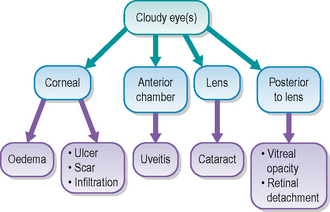
General clinical examination is usually unremarkable. On ocular examination the eye(s) appear slightly blue–grey in colour and the cornea appears slightly dull. Menace responses are usually normal as are pupillary light reflexes. The view of the iris and anterior chamber is hazy due to the presence of corneal oedema which manifests as a blue–grey mottled appearance. No ulceration is usually present (unless the disease has progressed to bullous keratopathy with ulceration – see the later section on prognosis).


The oedema can affect just one quadrant (e.g. dorsolaterally) or the entire cornea, and can be uni- or bilateral (but is not usually symmetrical – one eye will probably be more severely affected initially). The corneal oedema outlines the shape of the cornea and this gives the false impression that the eye is ‘bulging’. Normally the cornea is completely transparent and its convex shape is not apparent – one sees the flat iris instead – both from in front and laterally. Once the outline is visible due to the oedema then the shape is more obvious and the appearance is of the eye bulging. This must be differentiated from globe enlargement due to glaucoma or exophthalmos causing the eye to be placed more anterior than usual. The corneal oedema gives a mottled appearance and by gently pressing the globe over the sclera (through the upper lids for example) the oedema becomes denser. Palpation in this way is unlikely to be painful – whereas with a case of glaucoma or severe uveitis causing the oedema the patient would resent such examination due to the discomfort this would cause. Conjunctival hyperaemia can be present but is normally only very mild, providing ulceration is not a feature. Similarly, no significant ocular discharge is normally present.


Intraocular examination is unremarkable – details might be hazy but the use of distant direct ophthalmoscopy will help to determine whether the lens is clear or is cataractous, while indirect ophthalmoscopy will assist in evaluating the fundus.


Sep 10, 2016 | Posted by in SMALL ANIMAL | Comments Off on Corneal endothelial dystrophy

Full access? Get Clinical Tree

Get Clinical Tree app for offline access