Chapter 5 Cornea
INTRODUCTION
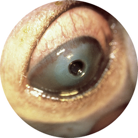
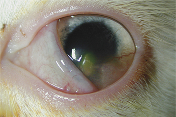
Figure 5-32 Descemetocele in a dog. The base of the ulcer looks black because the image appears against the dark iris. If the photograph had been taken against the tapetal reflex, the image would have resembled Figure 5-33. Corneal neovascularization is present.
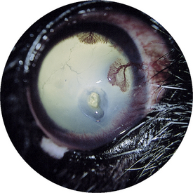
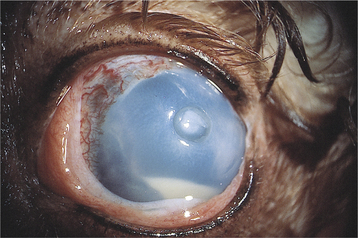
Figure 5-37 Chronic keratoconjunctivitis sicca (KCS) with healing deep corneal ulcer (see staining pattern in Figure 5-38).
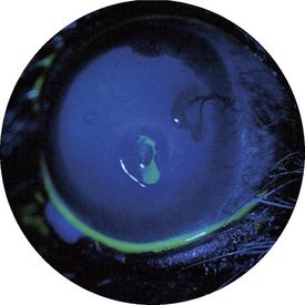
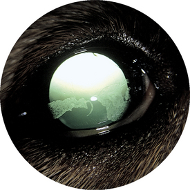
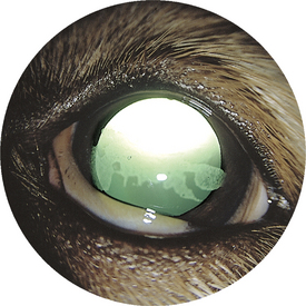
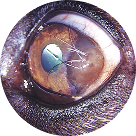
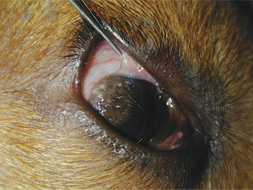
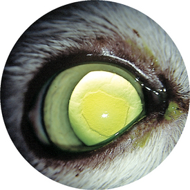
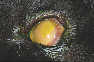
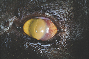
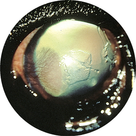
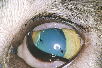
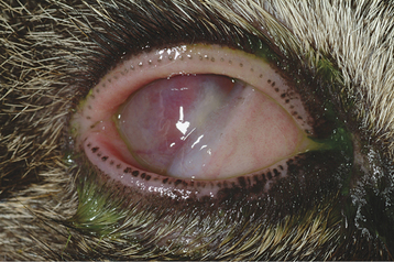
Figure 5-10 Superficial nonhealing corneal ulcer in a cat. Note the loose edges of epithelium (arrows).
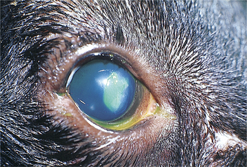
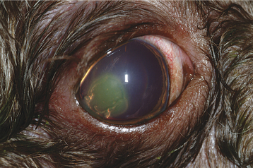
Figure 5-14 Nonhealing superficial ulcer. Note the loose tags of epithelium at the edge of the ulcer.
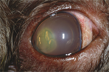
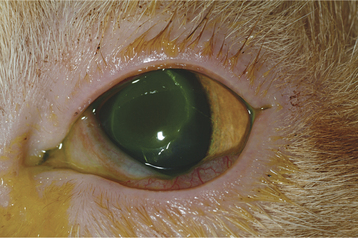
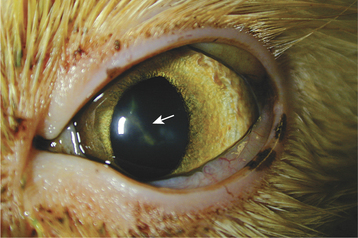
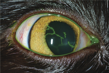
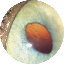
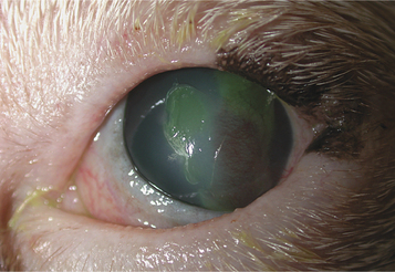
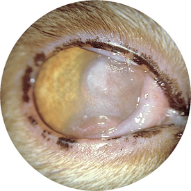
Figure 5-20 Feline herpesvirus keratitis. Corneal ulceration, edema, and vascularization are present.
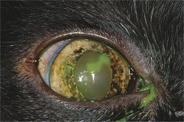
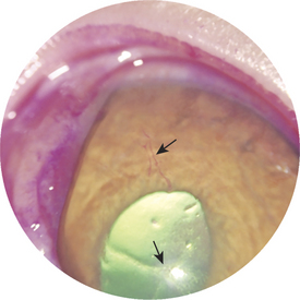
Figure 5-22 Herpesvirus keratitis stained with rose bengal in a cat. Faint, linear, red-stained lesions can be seen.
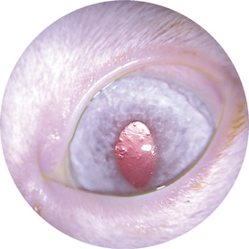
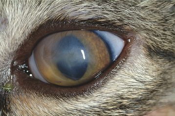
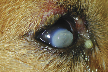
Figure 5-24 Corneal scarring in a cat undergoing treatment with trifluridine and oral lysine for herpesvirus infection.
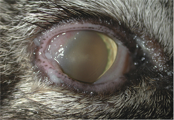
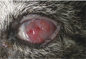
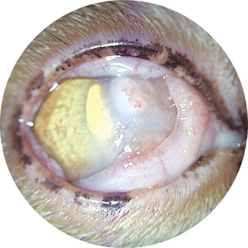
Figure 5-26 Neovascularization and cellular infiltrate in the cornea and nictitans of a cat with eosinophilic keratitis.
< div class='tao-gold-member'>
Stay updated, free articles. Join our Telegram channel

Full access? Get Clinical Tree


