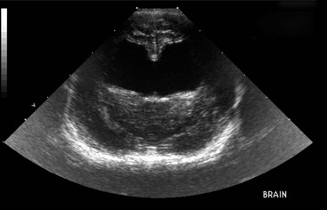
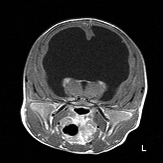
Congenital Hydrocephalus

Chapter 225


Congenital Hydrocephalus


Causes


Hydrocephalus is active distention of the ventricular system of the brain caused by obstruction of the flow of cerebrospinal fluid (CSF) from its point of production to its point of absorption (Rekate, 2009). CSF is produced at a constant rate by the choroid plexuses of the lateral, third, and fourth ventricles; the ependymal lining of the ventricular system; and blood vessels in the subarachnoid space. The CSF circulates through the ventricular system into the subarachnoid space, where it is absorbed by arachnoid villi. Obstruction can be caused by developmental abnormalities or acquired lesions such as neoplasia or inflammatory lesions.


A number of conditions such as infarction and necrosis can result in decreased volume of brain parenchyma that leaves a vacant space filled passively with CSF. Although this situation previously was referred to as hydrocephalus ex vacuo, it does not cause active distention of the ventricles and therefore is not classified as hydrocephalus.


Early hydrocephalus initially damages the ependymal lining of the ventricles. This allows water and larger molecules to leak into the adjacent white matter, causing periventricular edema. Further enlargement of the ventricles compresses the white matter, which leads to demyelination and axonal degeneration. The septum pellucidum separating the lateral ventricles can become fenestrated or completely destroyed, so that one single, large ventricle is created (Figure 225-1). In some cases, the cerebral cortex is preserved. In more severe cases, the cortex becomes thin, with neuronal vacuolation and loss of neurons. This affects the prognosis after surgical shunting. If the cortex is preserved, shunting results in reexpansion of the white matter and regeneration of remaining axons. However, if the cortex is damaged, the neuronal damage persists even after shunting. With severe obstruction to CSF flow, the volume of CSF can increase so fast that it causes an increase in intracranial pressure, which leads to further brain damage and impairs blood flow to the brain.


Clinical Features


Based on the age of onset, hydrocephalus can be classified broadly as pediatric or acquired. Pediatric hydrocephalus is caused by developmental abnormalities, and clinical signs often are noticed by several months of age. Toy and brachycephalic dogs are at increased risk, including the Maltese terrier, Yorkshire terrier, English bulldog, Chihuahua, Lhasa apso, Pomeranian, toy poodle, cairn terrier, Boston terrier, pug, and Pekingese. The most commonly identified cause in these breeds is stenosis of the mesencephalic aqueduct associated with fusion of the rostral colliculi. In many cases, however, an obvious site of obstruction is not apparent. These cases may be due to obstruction at the level of the subarachnoid space or arachnoid villi, which is difficult to detect. Another possibility is the occurrence of intraventricular obstruction during a critical stage of development with later resolution of the obstructive lesion so that only the ventricular enlargement remains. Pediatric hydrocephalus also may be associated with other malformations such as meningomyelocele, Chiari’s malformation, Dandy-Walker syndrome, and cerebellar hypoplasia. Many toy and brachycephalic dogs have enlarged ventricles with no apparent neurologic dysfunction. Hydrocephalus is seen sporadically in kittens, and although a genetic basis has been suggested in the Siamese, this has not been confirmed with genetic studies.


Clinical signs of pediatric hydrocephalus include an enlarged, dome-shaped head with persistent fontanelles and open cranial sutures. However, not all patients with a persistent fontanelle have hydrocephalus, and not all patients with pediatric hydrocephalus have a persistent fontanelle. Enlargement of the calvaria can be assessed subjectively by noticing whether the most lateral aspect of the parietal bone extends laterally beyond the level of the zygomatic arch. There may be ventral or ventrolateral strabismus due to either malformation of the orbit or brainstem dysfunction.


Affected patients often are unthrifty and smaller than normal. Common neurologic deficits include abnormal behavior and cognitive dysfunction, such as inability to become house trained. Visual deficits include unilateral or bilateral blindness with normal pupillary function. (One should remember that the menace response may not develop until at least 4 weeks of age in normal puppies and kittens.) Ataxia, seizures, circling, and vestibular dysfunction also are possible.


The clinical course is variable and difficult to predict. Neurologic deficits can progress over time, remain static, or even improve after 1 to 2 years of age. The condition of affected patients often is fragile and can worsen later in life coincident with other diseases. Patients with very large lateral ventricles and a thin cerebral cortex are at risk of intracranial hemorrhage from relatively trivial head trauma that results in tearing of bridging veins. This can result in chronic subclinical hematomas or sudden neurologic deterioration due to intracranial bleeding.


Acquired hydrocephalus can develop at any age and can be caused by neoplasia, head trauma, and meningoencephalitis. Neurologic deficits are similar to those in young animals, but if hydrocephalus develops after the cranial sutures have closed, malformation of the skull does not develop. Clinical signs also may reflect the underlying cause of the hydrocephalus.


Diagnosis


Diagnosis is based on the clinical features and brain imaging to assess ventricular size and identify any specific causes. Ventricular size usually is assessed subjectively by noting the progressively greater proportion of the intracranial volume occupied by the lateral, third, or fourth ventricles. However, there is poor correlation between clinical signs and ventricular size. Also, symmetric or asymmetric enlargement of the lateral ventricles is relatively common in normal animals. Therefore diagnosis of hydrocephalus is based on clinical features, not just ventricular size.


In pediatric patients with persistent fontanelles, ultrasonography allows easy detection of obviously enlarged ventricles. Normal-sized ventricles appear as paired, slitlike anechoic structures just ventral to the longitudinal fissure on either side of the midline. Enlarged ventricles are seen easily as paired anechoic regions. With marked ventricular enlargement, the septum pellucidum that normally separates the lateral ventricles is absent and the ventricles appear as a single, large anechoic structure (Figure 225-2).


< div class='tao-gold-member'>

Stay updated, free articles. Join our Telegram channel

Jul 18, 2016 | Posted by in PHARMACOLOGY, TOXICOLOGY & THERAPEUTICS | Comments Off on Congenital Hydrocephalus

Full access? Get Clinical Tree

Get Clinical Tree app for offline access