Chapter 12 Congenital Heart Disease
INTRODUCTION
Incidence
• The most common congenital heart defects in dogs include patent ductus arteriosus (PDA), pulmonic stenosis (PS), aortic stenosis, ventricular septal defect (VSD), and tetralogy of Fallot (Box 12-1). Several breed predispositions have been identified (see Appendix 1: Canine Breed Predilections for Heart Disease).
Box 12-1 Classification of Congenital Defects According to Pathophysiology
| CANINE | FELINE |
|---|---|
| Defects primarily causing volume overload | Defects primarily causing volume overload |
| Systemic to pulmonary (left-to-right) shunting | |
| Common | Common |
| Patent ductus arteriosus | Ventricular septal defect |
| Ventricular septal defect | Patent ductus arteriosus |
| Uncommon | Atrial septal defect |
| Atrial septal defect | Endocardial cushion defect |
| Endocardial cushion defect | Uncommon |
| (Pseudo) truncus arteriosus | Truncus arteriosus |
| Valvular regurgitation | Valvular regurgitation |
| Common | Common |
| Mitral dysplasia | Mitral dysplasia |
| Tricuspid dysplasia | Tricuspid dysplasia |
| Uncommon | |
| Pulmonic insufficiency | |
| Aortic insufficiency | |
| Defects primarily causing pressure overload | Defects primarily causing pressure overload |
| Common | Common |
| Pulmonic stenosis | Dynamic subaortic stenosis |
| Subaortic stenosis | Uncommon |
| Uncommon | Pulmonic stenosis |
| Valvular aortic stenosis | Pulmonary artery branch stenosis |
| Coarctation and interruption of the aorta | Fixed subaortic stenosis |
| Cor triatriatum dexter | Valvular aortic stenosis |
| Cor triatriatum dexter | |
| Cor triatriatum sinister | |
| Defects primarily causing cyanosis | Defects primarily causing cyanosis |
| Common | Common |
| Tetralogy of Fallot | Tetralogy of Fallot |
| Uncommon | Endocardial cushion defect |
| Pulmonary to systemic shunting (ventricular septal defect [VSD]) | Uncommon |
| Pulmonary to systemic shunting (patent ductus arteriosus [PDA]) | Pulmonary to systemic shunting (VSD) |
| Tricuspid atresia/right ventricular hypoplasia | Pulmonary to systemic shunting (PDA) |
| Double outlet right ventricle | Double outlet right ventricle |
| Transposition of the great vessels | Truncus arteriosus |
| Truncus arteriosus | |
| Aorticopulmonary window | |
| Miscellaneous cardiac and vascular defects | Miscellaneous cardiac and vascular defects |
| Common | Common |
| Peritoneopericardial diapharagmatic hernia | Peritoneopericardial diapharagmatic hernia |
| Persistent right aortic arch | Endocardial fibroelastosis |
| Persistent left cranial vena cava | Uncommon |
| Uncommon | Persistent right aortic arch |
| Endocardial fibroelastosis | |
| Pericardial defects | |
| Anomalous pulmonary venous return | |
| Double aortic arch | |
| Retroesophageal left subclavian artery | |
| Situs inversus |
Diagnosis
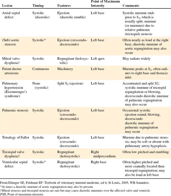
• Generally, a congenital heart defect is suspected when a heart murmur is detected in a young dog or cat (Table 12-1).
• A tentative diagnosis can be made based on results of a complete cardiovascular physical examination, routine ECG, and radiography (see Table 12-1).
Therapy
• Advances since the early 1990s allow for the treatment of certain stenotic lesions by balloon valvuloplasty, a technique employing a small balloon located at the end of a cardiac catheter, and transcatheter occlusion of PDA using a Gianturco coil or similar device. Surgical correction or palliation is possible for certain defects.
SPECIFIC DEFECTS
Pathophysiology
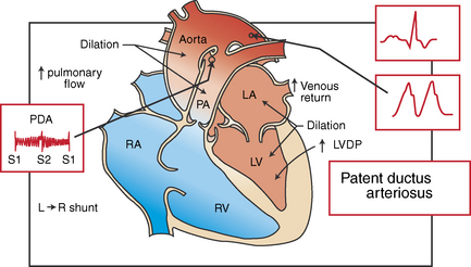
• When pulmonary vascular resistance is normal, blood continually shunts from the aorta (high resistance) into the pulmonary circulation (low resistance). Shunting in this fashion (systemic to pulmonary) is referred to as left-to-right and represents the most common pattern in PDA (Figure 12-1).
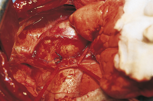
• Failure of ductal closure is due to an abnormal amount of elastic fibers compared to contractile smooth muscle fibers (so-called extension of the noncontractile wall structure of the aorta into the ductus arteriosus). Varying amounts of normal smooth muscle causes varying degrees of ductal closure; this creates a structure that ranges from a funnel-shaped ductus that narrows on the pulmonary artery side of the ductus (Figure 12-2) to a tubelike structure without narrowing.
Diagnosis
Etiology and Breed Disposition
• Breeds predisposed or at increased risk include Bichon Frisé, Chihuahua, Cocker Spaniel, Collie, English Springer Spaniel, German Shepherd, Keeshond, Labrador Retriever, Maltese, Newfoundland, Poodle, Pomeranian, Shetland Sheepdog, and Yorkshire Terrier.
Diagnostic Testing
Thoracic Radiographs
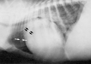
• The radiographic signs of PDA (Figures 12-3 and 12-4) vary considerably with the volume of blood being shunted, the age of the animal, and the degree of cardiac decompensation.
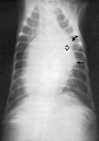
Figure 12-4 Patent ductus arteriosus (dorsoventral projection) in a 3-month-old poodle. There is generalized heart enlargement, an aneurysm-like bulge of the descending aorta (open arrow), a bulge of the main pulmonary artery (curved arrow), and enlargement of the left auricle (straight arrow). Alveolar infiltrate due to pulmonary edema is present in the caudal lung lobes.
Selective Angiocardiography
• The diagnosis of a left-to-right PDA can be made by injecting contrast media into the aortic arch. The simultaneous filling of the main pulmonary artery and the aorta is diagnostic for PDA. Likewise, a main pulmonary artery contrast injection can be used to confirm the diagnosis of a right-to-left shunt (simultaneous filling of main pulmonary artery and aorta).
Differential Diagnosis
Natural History

