CHAPTER | 10 Congenital Diseases
Epidermolysis Bullosa
Treatment and Prognosis
2 Trauma should be avoided by keeping the affected animal indoors, away from other animals, and handling it carefully.
3 Appropriate systemic antibiotics should be administered as needed for secondary bacterial infection.
4 The prognosis for severely affected animals is poor. With proper environmental management, mildly affected animals may enjoy a reasonable quality of life. Affected animals should not be bred.
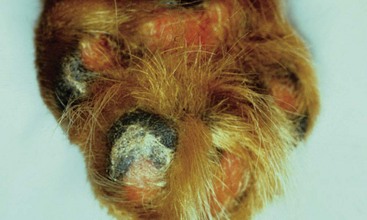
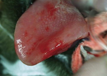
FIGURE 10-2 Epidermolysis Bullosa.
Sloughing of the footpads. The superficial epidermis is peeling off.
(Courtesy P. Rakich.)
Familial Canine Dermatomyositis
Diagnosis
2 Dermatohistopathology (may be nondiagnostic): scattered epidermal basal cell degeneration; perifollicular inflammatory infiltrates of lymphocytes, histiocytes, and variable numbers of mast cells and neutrophils; follicular basal cell degeneration; and follicular atrophy are highly suggestive findings but may not be present, especially in chronic or scarred lesions.
3 Electromyography: fibrillation potentials, bizarre high-frequency discharges, and sharp waves are seen in affected muscles.
Treatment and Prognosis
4 Intact females should be spayed because estrus, pregnancy, and lactation exacerbate the disease. Affected males should be neutered so they cannot reproduce.
5 Daily supplementation with oral essential fatty acids and treatment with vitamin E 400 to 800 IU PO every 24 hours may be beneficial for the skin lesions. Improvement should be seen after 2 to 3 months of therapy (see Table 8-2).
6 Treatment with pentoxifylline (Trental) 25 mg/kg PO every 12 hours with food may be beneficial in some dogs. Improvement should be seen within 1 to 3 months of therapy.
7 Cyclosporine (Atopica) 5–10 mg/kg PO administered every 24 hours may be beneficial (effects are seen in approximately 4–6 weeks). Then, dosage frequency should be tapered down to every 48 to 72 hours. Glucocorticoids can be used initially to speed response. As of this writing, no statistically significant increase in tumor risk or severe infection is known to result from the immune effects of cyclosporine.
8 Prednisone 1 mg/kg PO every 24 hours until lesions improve (approximately 7–10 days), then tapered off, may be used for acute flare-ups; however, prolonged steroid usage may exacerbate muscle atrophy.
9 The prognosis is variable, depending on the severity of the disease. Skin lesions in minimally affected dogs tend to resolve spontaneously, with no scarring. Skin lesions in mildly to moderately affected dogs usually resolve eventually, but residual scarring is common. Even when lesions resolve, however, relapses may occur later on, when the dog is an adult. In severely affected dogs, dermatitis and myositis do not resolve, and the prognosis for long-term survival is poor. Regardless of disease severity, affected dogs should not be bred.
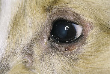
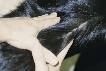
FIGURE 10-5 Familial Canine Dermatomyositis.
The same dog as in Figure 10-4. Active lesions have resolved, leaving alopecic, scarred skin.
(Courtesy M. Mahaffey.)
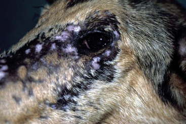
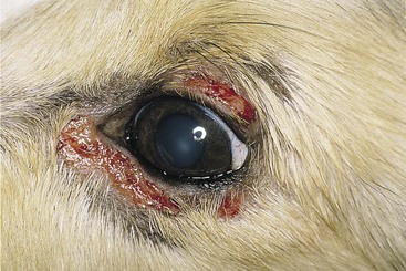
FIGURE 10-6 Familial Canine Dermatomyositis.
Alopecia and scarring on the face of an adult collie. The erythematous macules were active lesions.
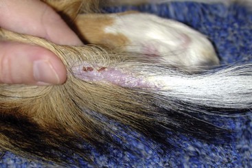
FIGURE 10-7 Familial Canine Dermatomyositis.
Alopecic, erythematous, crusting dermatitis on the tail of a collie with dermatomyositis.
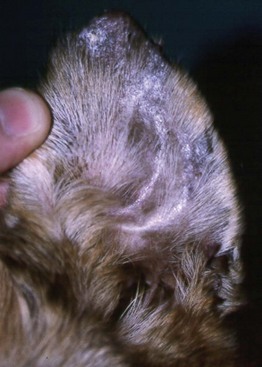
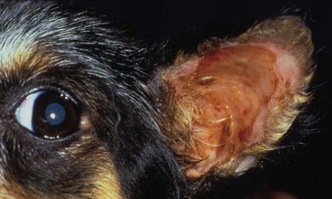
FIGURE 10-9 Familial Canine Dermatomyositis.
Crusting, erosive lesions on the ear pinna of an adult collie with chronic lesions.
Stay updated, free articles. Join our Telegram channel

Full access? Get Clinical Tree


