CHAPTER 126 Collateral Desmitis of the Coffin Joint
Collateral desmitis of the coffin joint has long been ignored as a potential cause of foot lameness in horses because of practitioners’ inability to image the collateral ligament with radiography and the absence of telling clinical signs. First ultrasonography—and later magnetic resonance imaging (MRI)—have focused attention on this important structure and its appearance in health and disease. The frequency with which collateral desmitis has been diagnosed has therefore increased dramatically in recent years. In one study it was considered to be the second most important single injury responsible for foot lameness in horses without radiographic abnormalities (behind digital deep digital flexor tendonitis), whereas in another study it was found to be the most common injury in Warmblood sport horses.
CLINICAL EXAMINATION
Visual Inspection and Palpation
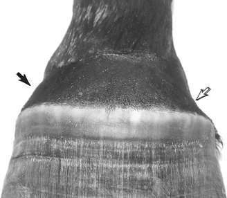
Most horses with collateral desmitis or enthesopathy do not have localizing signs. Distension of the dorsal pouch of the coffin joint may be detected but is not a characteristic finding. A discrete, palpable swelling may occasionally be found at the level of the origin of one of the ligaments, immediately proximal to the dorsomedial or dorsolateral aspect of the coronary band (Figure 126-1). The medial collateral ligament is injured nearly twice as often as the lateral. In a small proportion of horses, both lateral and medial ligaments are affected.
Stress Tests
Some investigators report that a pain response cannot be induced by passive manipulation of the digit in horses with collateral desmitis of the coffin joint. In the opinion of the second author of this chapter, however, horses with collateral desmitis are likely to react painfully to digit flexion performed with the examiner pulling on the toe of the hoof capsule while facing caudally. Horses with collateral enthesopathy are less likely to resent digit flexion. The second author further believes that a positive flexion test is a favorable prognostic sign in horses with collateral ligament injury because it reflects minimal or no involvement of the attachment sites of the ligament. Monitoring the pain response to digit flexion is also useful in deciding when a horse with collateral desmitis may return to training.
Regional Anesthesia
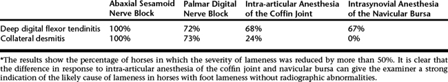
Not all horses suspected of having soft tissue injury in the foot have the benefit of undergoing MRI examination. In these instances, the response to different anesthetic techniques in the foot can help to distinguish between the two most common soft tissue injuries, collateral desmitis and digital tendonitis of the deep digital flexor tendon (Table 126-1).