Chapter 83: Canine Brucellosis

Web Chapter 83


Canine Brucellosis


Brucellosis is the primary infectious venereal disease of concern in canine reproduction. Brucella canis causes reproductive failure in both the male and female dog. Screening for B. canis is an important part of the prebreeding evaluation of any dog and should be included in the initial diagnostic approach to any case of canine abortion or infertility. Because the incidence of canine brucellosis is low in many parts of the world, including the United States, breeder compliance with regular testing can wane, which makes continued veterinary vigilance important. Many experts consider canine brucellosis to be an incurable, potentially zoonotic disease. Canine brucellosis can affect neutered dogs and bitches as well as individuals that have not been bred.


Etiology and Microbiology


Canine brucellosis is caused by B. canis, a small gram-negative, non–spore forming aerobic coccobacillus. Brucella abortus, Brucella melitensis, and Brucella suis occasionally have caused canine infections but comparatively are very rare. Transmission occurs through direct exposure to bodily fluids (semen, lochia, aborted fetuses or placentas, milk, and urine) containing an infectious dose of organisms. A large number of organisms are shed in the vulvar discharge of bitches 4 to 6 weeks after abortion. The highest concentration of organisms is found in the semen of infected dogs 2 to 3 months after infection, with lesser numbers of the bacteria shed for years. Urine can serve as a contaminated vehicle because of the proximity of the urinary and genital tracts in the dog. Milk also can serve as a vehicle for shedding. Therefore transmission is primarily oral (i.e., through the mucous membranes) and often is associated with the ingestion of infectious materials. Urine is important in transmission when groups of dogs are housed together. The aerosol route is important in kennel environments because of nasal and conjunctival exposure. Venereal transmission occurs, but outbreaks more commonly are associated with nonvenereal mucous membrane transmission. B. canis is short lived outside the dog and is inactivated readily by common disinfectants such as 1% sodium hypochlorite, 70% ethanol, iodine-alcohol solutions, glutaraldehyde, and formaldehyde.


Brucella organisms attach to and penetrate the mucous membranes; virulence is proportional to the infectious dose. After replication in regional lymph nodes, bacteremia occurs within 7 to 30 days after exposure with subsequent transportation to monocyte-macrophage series cells, bone marrow, spleen, prostate, uterus, and placenta. B. canis has a predilection for steroid-dependent (reproductive) tissues. It survives facultatively within monocytes and macrophages. Brucella organisms are able to survive and multiply in monocyte-macrophage cells because they inhibit the bactericidal myeloperoxidase-peroxide-halide system by releasing 5′-guanosine and adenine. Early in infection polymorphonuclear cells and macrophages are relatively ineffectual in killing B. canis intracellularly.


Brucella organisms can be identified in the rough endoplastic reticulum of placental trophoblastic giant cells in an infected bitch’s gravid uterus. Severe necrotizing placentitis with infarction of the labyrinth region, coagulation necrosis of the chorionic villi, and necrotizing arteritis result in fetal death. The organism can be found in the gastric contents of the aborted fetuses. Necrotizing vasculitis causing granulomatous lesions results in epididymal and subsequent testicular and prostatic abnormalities.


Epizootiology


Members of the Canidae family are the natural hosts of B. canis, and any mature, reproductively active dog of any breed is susceptible to infection. Canine brucellosis occurs most commonly as an outbreak in a large commercial kennel and less commonly in privately owned dogs. Outbreaks of canine brucellosis traditionally have a geographic component; incidence is higher in the southernmost states of the United States, and the disease is more common in Mexico, Central and South America, the People’s Republic of China, and Japan. The incidence in Europe has been low. The increased practice and success of canine semen processing for exportation (chilling and cryopreservation) for the purpose of artificial insemination now makes canine brucellosis a concern worldwide since direct mucosal or venereal contact among dogs is no longer necessary for transmission.


Humans can become infected with B. canis, although apparently rarely. Approximately 40 cases of human infection have been reported in several countries; however, the actual number is unknown since human cases rarely are diagnosed or, if diagnosed, reported. Transmission to humans most commonly occurs through contact with semen from an infected dog, vulvar discharge from an infected bitch, aborted fetuses or placentas, or direct accidental laboratory exposure.


Clinical Signs


Canine brucellosis has high morbidity but low mortality. The clinical systemic signs often are subtle and can include suboptimal athletic performance, lumbar pain, lameness, weight loss, and lethargy.


The primary clinical sign of brucellosis in the bitch is pregnancy loss, which can occur early in gestation (20 days), resulting in fetal resorption, or more commonly (approximately 75% of cases) later in gestation (generally 45 to 59 days), resulting in abortion. Bitches with pregnancy loss early in gestation can appear to be infertile unless early ultrasonographic pregnancy evaluation is performed. Rarely, a litter is carried to term in an infected bitch; neonates die a few days after birth. Nongravid bitches can be infected subclinically or can show regional lymphadenopathy (pharyngeal if orally acquired, inguinal and pelvic if venereally acquired).


The primary acute clinical signs of brucellosis in the male dog reflect disease of the portions of the reproductive tract participating in the maturation, transport, and storage of spermatozoa. Epididymitis is common, with associated orchitis, scrotal dermatitis, and resultant deterioration of semen quality and fertility. Over the long term, testicular atrophy and infertility can occur. The organism can be found in the prostate gland and urine. Antisperm antibodies develop in association with brucellosis-induced epididymal granulomas and further contribute to infertility. Diminished sperm counts (oligospermia), poor sperm motility (asthenospermia), and increased morphologic abnormalities (teratospermia) are characteristic of semen in a Brucella-infected dog. Detached sperm heads, proximal and distal cytoplasmic droplets, and acrosomal deformities are the most common morphologic abnormalities. Sperm head-to-head agglutination suggests the presence of antisperm antibodies. Pyospermia develops 3 to 4 months after infection. An absence of sperm in the ejaculate (azoospermia) also can result.


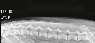
Chronic infections in either sex can result in uveitis, granulomatous splenitis, discospondylitis (Web Figures 83-1, and 83-2), granulomatous dermatitis, meningoencephalitis, and nephritis. Bacteremia can persist for years, and asymptomatic dogs can remain infectious for long intervals.


< div class='tao-gold-member'>

Stay updated, free articles. Join our Telegram channel

Jul 18, 2016 | Posted by in PHARMACOLOGY, TOXICOLOGY & THERAPEUTICS | Comments Off on Chapter 83: Canine Brucellosis

Full access? Get Clinical Tree

Get Clinical Tree app for offline access