Chapter 80: The Neuro-ophthalmic Examination

Web Chapter 80


Anisocoria and Abnormalities of the Pupillary Light Reflex


The Neuro-ophthalmic Examination


The neuro-ophthalmic examination is a tool that localizes a defect to the nervous system or the eye. The ultimate goal is to provide the information necessary to proceed with diagnosis, prognosis, and treatment of the pet. This chapter summarizes key aspects of this examination and interpretation of the findings.


Anisocoria (unequal size of the pupils) is abnormal and is a common neuro-ophthalmic problem. This abnormality also may be associated with abnormalities of the pupillary light reflex (PLR). The clinician’s role is to determine which pupil is abnormal and whether the underlying cause is ophthalmic or neurologic. This chapter provides a logical guide to address frequent owner concerns: What is wrong? What caused it? How is it treated? Will it get better? These goals are best achieved by first examining the eye with the normal pupil, assessing the PLRs, and then evaluating the eye with the abnormal pupil. Identification of lesions enables a short list of potential causes to be assessed. Once the cause is known, a prognosis can be given.


There are two types of anisocoria: static and dynamic. Static anisocoria is unequal pupils when both eyes are receiving equal illumination. In a patient with dynamic anisocoria, the difference in pupil size depends on the stimulation of one pupil. Some degree of dynamic anisocoria is normal because of incomplete decussation of afferent fibers at two locations: the optic chiasm and the pretectal nucleus. At the optic chiasm, nerve fibers cross to the contralateral side, whereas at the pretectal nucleus, nerve fibers cross back to the original (ipsilateral) stimulated side. Although pupils of both eyes react to light when it is directed at only one eye, the iris sphincter muscle of the directly stimulated eye receives more efferent impulses than the nonstimulated eye. Therefore it is normal for the pupil receiving direct stimulation to be smaller than the fellow (nonstimulated) pupil. This is termed physiologic dynamic anisocoria and does not indicate a pathologic condition.


For all patients in which a neuro-ophthalmic defect, anisocoria, or abnormal PLR is diagnosed, a thorough medical history that includes past medical conditions, current medications, current medical conditions, behavioral changes, and physical signs should be elicited. The owner should be asked about any trauma or absence of the pet from its environment, as well as prior treatment by either the owner or a previous veterinarian. For example, if atropine is used as a mydriatic instead of a more appropriate short-acting agent such as tropicamide, the resulting mydriasis could last up to 2 weeks. Alternatively, owners may try home remedies, which can include medications used for a previous condition, that may alter the pupil size. Behavioral changes often accompany visual deficits because the pathways for both are intimately associated in the brain, particularly the forebrain.


A complete ophthalmic examination (see Chapter 242), including measurement of intraocular pressure, examination of the cranial nerves, and a full physical examination, always should be done. Recording all examination findings is essential (Web Figure 80-1) to compile the information that must be reviewed before making a decision regarding the cause of the neuro-ophthalmic defect or anisocoria.


Role of the Cranial Nerves in the Neuro-ophthalmic Examination


Most of the cranial nerves are involved in vision, PLRs, eye position, and movement. Cranial nerve II (CN II), or the optic nerve, is the afferent arm of the PLR and vision and transmits sensory information to the brain. Parasympathetic fibers of CN III, the oculomotor nerve, comprise the efferent arm of the PLR. CN III also innervates the levator palpebrae superioris muscle (to elevate the upper eyelid) and most of the extraocular muscles. CN IV, the trochlear nerve, innervates the dorsal oblique muscle that passes over a trochlea to rotate the globe medially. CN V, the trigeminal nerve, provides sensory input via the ophthalmic (medial) and maxillary (lateral) branches to the surfaces of the eye (cornea, conjunctiva, periocular skin) and provides afferent input to the lacrimal gland. CN VI, the abducens nerve, serves the lateral rectus muscle (to abduct the globe) and the retractor bulbi muscles. A helpful acronym for remembering the innervation of the extraocular muscles is LR6, SO4, similar to a chemical equation. This denotes that the lateral rectus and the retractor bulbi are innervated by cranial nerve 6 and the superior oblique by cranial nerve 4 (whereas all other extraocular muscles are controlled by CN III). CN VII, the facial nerve, innervates the facial muscles controlling eyelid closure, important in the menace and palpebral reflex. CN VIII, the vestibular nerve, is responsible for coordination of globe position and gaze.


Functional Anatomy of the Pupillary Light Reflex
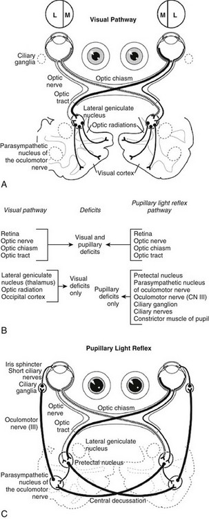

Pupil size (and shape in the cat) is determined by the balance between parasympathetic and sympathetic innervation of the iris. Although emotional and other non–neuro-ophthalmic factors can affect this aperture, pupil size depends in large part on the amount of light illuminating the retina and the afferent and efferent pathways of the PLR. An understanding of the anatomy of the PLR is essential (Web Figure 80-2, B and C), along with recognition that the PLR reflex is subcortical (without cerebral cortical involvement) and that the reflex is not an indicator of vision. The response of only a few photoreceptors is required for the PLR to occur.


The afferent arm of the PLR for each eye consists of the retina, optic nerve, optic chiasm, and optic tract. Nerve fibers originating in the medial retina decussate to the opposite optic tract, whereas fibers from the lateral retina remain in the ipsilateral tract. In the dog approximately 75% of the optic nerve fibers decussate to the contralateral optic tract. In the cat approximately 65% of the optic nerve fibers decussate. The optic tracts carry impulses beyond the decussation and include both sensory visual fibers and pupillomotor fibers that differ according to their destinations. This fact is useful for localizing lesions by noting the presence or absence of vision and PLR in each eye. The pupillomotor fibers progress to the pretectal nucleus, and from there the majority of these fibers decussate to the parasympathetic nucleus of the oculomotor nerve (previously known as the Edinger-Westphal nucleus) on the contralateral side. This second decussation marks a return to the side of the originating (light) impulse. In contrast, the visual fibers proceed from the optic tract sequentially to the ipsilateral lateral geniculate nucleus, the optic radiation, and the occipital (visual) cortex. Therefore lesions that cause anisocoria and affect both PLRs and vision can be localized to the retina, optic nerve, optic chiasm, or optic tract (see Web Figure 80-2). By contrast, lesions affecting vision, but not PLRs, are located in the lateral geniculate nucleus, optic radiation, or occipital cortex.


The efferent arm of the PLR consists of parasympathetic fibers in the oculomotor nerve (CN III) that travel to the ciliary ganglion, postganglionic ciliary nerves, and finally the iris sphincter muscle. In the dog there are five to eight short ciliary nerves that contain both sympathetic and parasympathetic efferent fibers and are considered “mixed.” Therefore a lesion of the ciliary nerve in the dog results in a circular, midrange pupil. By contrast, the cat has only two short ciliary nerves, the nasal (medial) and malar (lateral) nerves; these are solely parasympathetic. Thus lesions involving the feline nerves cause more dramatic pupil dilation than in the dog because sympathetic function is still present and is now unopposed. If only the medial or lateral branch is damaged, dyscoria (altered pupil shape) results in a D-shaped or reverse D–shaped pupil in addition to anisocoria.


The sympathetic fibers innervate the iris dilator muscle. The three-neuron pathway that the sympathetic axons traverse is more complex. The central fibers originate in the hypothalamus and exit the brain with the tectotegmentospinal tract. The preganglionic cell bodies lie in spinal cord segments T1 to T3 and issue fibers that travel with the ventral spinal nerve roots. These pass cranially via the thoracic and cervical sympathetic trunk to terminate in the cranial cervical ganglion, caudal and medial to the tympanic bulla. Postganglionic fibers then exit the ganglion and pass through the cavernous sinus into the periorbita, nasociliary nerve, long ciliary nerve, ciliary body, and finally the iris dilator muscle.


The Neuro-ophthalmic Examination


The neuro-ophthalmic examination has a number of components including general observations, neurologic evaluation, assessment of the pupils and PLR, and general ophthalmic examination.


Observation of Behavior, Gait, and Facial Symmetry and Assessment of Cranial Nerves


The examination begins as the patient is walking toward the examination room. Does the pet have a normal gait or are there abnormalities such as ataxia, hugging the wall, leading with the nose, or tentativeness in movement? Is there a head tilt or circling? When the pet is off leash in the examination room, does it explore (an indication of a sighted or confident long-blind dog) or does it stay close to its owner? If it explores, does it bump into things? Does the pet tend to “lead with one eye” while bumping into objects on the other side?


A crucial part of the neuro-ophthalmic examination is observation of the carriage of the head and assessment of facial and ocular symmetry. It is quite easy to miss neurologic lesions by focusing on only the apparently affected eye in the assessment, especially in cases of anisocoria. For example, Horner’s syndrome and facial nerve paralysis are recognized most often during the initial observation of facial symmetry.


CN II to VIII should be assessed during the neuro-ophthalmic examination. Elicitation of the menace response tests CN II and VII. Pupil size and PLR are observed in order to assess CN II and parasympathetic fibers of CN III along with the sympathetic innervation. The size of the eyelid opening is affected by CN III and VII and sympathetic nerves. The palpebral reflex is determined by CN V and VII. The position of the eyes is controlled by CN III, IV, and VI, the vestibular system, and the brainstem. The oculocephalic reflex induces nystagmus and tests CN VIII along with CN III, IV, VI, and a relatively long segment of brainstem connections from the pontomedullary junction to the midbrain.


Assessment of Visual Status


It is important to determine the visual status of each eye before checking the PLRs. Each eye should be assessed individually because the patient can easily compensate for unilateral blindness. The menace response usually is more notable during the initial part of the examination, before the pet has been subjected to bright lights shone in its eyes but after it is relatively calm on the table.


A properly performed menace test, done without creating air currents or noise, is a helpful test of vision. The menace test involves a learned response rather than a reflex and is not developed in small animals until 8 to 12 weeks of age. The appropriate response to this gesture is to close the eyes rapidly. Frightened dogs and recalcitrant cats may not blink, even though visual. Often, a cat’s menace response is the slightest flicker of its eyelids. The pathway being tested is optic nerve→optic chiasm→optic tract→lateral geniculate nucleus→optic radiation→visual cortex→motor cortex→internal capsule and crus cerebri→the facial nuclei in the medulla→facial nerve→eyelid muscles. One hand should cover one eye as the other eye is being tested. The menacing gesture should appear suddenly in the pet’s visual field. An easy way to do this is to begin the gesture below the level of the pet’s eyes and rapidly bring the menacing hand straight up. Flicking the fingers toward the pet creates noise and air currents and therefore is unreliable. If the pet fails to respond, the reason could be that the facial nerve is not functioning, that the response has not been learned, that there is a cerebellar deficit, or that the examiner is not menacing enough! Facial nerve function should be checked by testing the palpebral reflex. If the eyelids close in response to being tapped, that response is intact. If only the globe retracts and the nictitans protrudes during the menace response, the facial nerve is not functioning, but the pet is visual. Note that the trigeminal nerve (CN V) provides the afferent arm of the palpebral reflex via ocular and periocular sensation and also must be assessed; however, facial nerve problems are more common than trigeminal disorders. If the facial nerve is intact and the pet fails to respond, the examiner should gently tap the head to indicate that he or she will follow through with the menacing gesture, then repeat the menacing gesture without touching the head.


Observing an animal tracking cotton balls dropped in front of it while shielding one eye perhaps is a more reliable test of visual function. Tracking a laser pointer or the light from a penlight are good tests of vision in small animals, especially in cats, because they are notoriously reluctant to participate in the menace response.


Maze tests can be set up easily in the examination room using available objects such as chairs and wastebaskets. The pet should be taken to the opposite side of the room from the owner, should be turned to face away from the owner, and then should be called by the owner. This test can be done under photopic (normal room lighting) and scotopic (darkened room) conditions to test for subtle visual deficits, such as early progressive retinal atrophy.


Visual placing is an additional vision test performed by holding the pet under one’s arm as it is moved toward a table. The desired response is for the pet to lift the front limb and place it on the table before touching the table.


The dazzle reflex is a subcortical reflex performed by stimulating one eye with a bright light, with the expected result being closure of the eyelids. The pathway is CN II→rostral colliculus→CN VII. Unlike the menace response, the dazzle reflex still occurs in animals blind from cerebrocortical disease because the cerebrum is not involved in this pathway. This reflex also is present in animals that are blind due to a complete cataract, for example, but have an intact CN II. The response is absent in patients blind due to a subcortical lesion, such as glaucoma causing destruction of the optic nerve.

< div class='tao-gold-member'>

Stay updated, free articles. Join our Telegram channel

Jul 18, 2016 | Posted by in PHARMACOLOGY, TOXICOLOGY & THERAPEUTICS | Comments Off on Chapter 80: The Neuro-ophthalmic Examination

Full access? Get Clinical Tree

Get Clinical Tree app for offline access