Chapter 76: Canine Retinal Detachment

Web Chapter 76


Canine Retinal Detachment


Pathogenesis
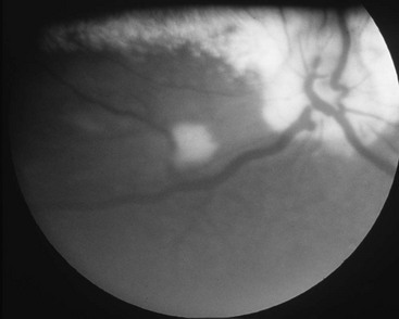

A retinal detachment (RD) is a separation of the neurosensory retina from the retinal pigment epithelium. In the normal eye the vitreous, retina, and lens are connected by various physical and chemical mechanisms. RDs may be caused by one of three main mechanisms: exudation (nonrhegmatogenous), retinal tears (rhegmatogenous), or traction pulling on the retina. Complicated RDs are those that involve more than one of these mechanisms (e.g., a retinal tear or hole that results from a vitreous traction band).


In small animals most RDs are exudative. The subretinal fluid usually is inflammatory but can be serous in diseases such as systemic hypertension (see Chapter 169). The subretinal fluid typically results from breakdown of the blood-ocular barrier in the retinal and choroidal vasculature. Because the choroidal vascular bed is much larger than the retinal vascular supply, a large amount of subretinal fluid usually indicates diffuse choroidal involvement, as seen in diseases such as chorioretinitis, systemic hypertension, or hyperviscosity. Initially areas of chorioretinitis appear as variably sized focal or multifocal areas of retinal elevation with indistinct borders. These active areas of chorioretinitis alter the course of the overlying retinal blood vessels and obscure or blur the ophthalmoscopic view of the underlying retinal pigment epithelium or tapetum as shown in Web Figure 76-1.


Rhegmatogenous RD is associated with the formation of one or more retinal tears or holes. This type of RD is less common than exudative RD in small animals and occurs more frequently in dogs than in cats. The pathogenesis of rhegmatogenous RD involves the presence of an abnormal retina (i.e., thinned as a result of degeneration, age, or other diseases) that is predisposed to formation of tears or holes, combined with an abnormal vitreous, as with vitreous syneresis (liquefaction), traction (from vitreal floaters or postinflammatory debris), or vitreous detachment. The vitreous gel is a homogeneous collagen fibril network with hyaluronic acid molecules filling the interfibrillar spaces. The vitreous is more firmly attached in three locations: the peripheral posterior lens capsule, the vitreous base that overlies the peripheral edge of the retina (the pars plana and ora ciliaris retinae), and the margin of the optic nerve. Since the vitreous hydrogel is attached to the lens and retina, perturbations of the vitreous (e.g., caused by inflammation or surgery) or of the lens can contribute to the development of rhegmatogenous RD by creating retinal traction, which can lead to tear or hole formation. Liquid from the vitreous then moves through the hole or tear into the subretinal space and exacerbates the RD.


Causes


Despite the multiple causes of RD, the following three features assist in differentiating or prioritizing the likely causes: (1) the type of RD (exudative, traction, or rhegmatogenous), (2) the nature of any subretinal fluid, and (3) whether the detachments are unilateral or bilateral. Although bilateral RD is strongly suggestive of systemic disease or congenital ocular disease, unilateral detachment does not rule out these causes. Serous subretinal fluid with or without hemorrhage is more common with hypertension, hyperviscosity syndromes, and rickettsial disease. When subretinal fluid is opaque from a marked cellular infiltrate, causes of exudative disease such as lymphosarcoma, idiopathic inflammatory uveodermatologic syndrome, and fungal infections should be considered more likely.


Ocular Anomalies


RD may be associated with severe retinal dysplasia, optic nerve colobomas, vitreous abnormalities, and retinal nonattachment (i.e., developmental failure of the two retinal layers to unite). Animals with RD present at or soon after birth and often have concurrent abnormalities such as microphthalmia or cataracts. Detachment may be unilateral or bilateral.


Later-onset genetic anomalies such as cataracts or vitreous degeneration may lead to RD. Rhegmatogenous RD also may occur because of vitreous liquefaction or cataract formation, particularly when the cataracts are rapidly forming or hypermature, or when there is lens-induced uveitis. Rhegmatogenous RD may occur as a sequela to cataract surgery (reported incidence, 1% to 4%) or lens luxation or subluxation. Many of the terrier breeds suffer from genetically based primary lens luxation, and retinal tearing and detachment may occur with or without lens surgery in these breeds. Premature and severe vitreal degeneration occurs in the Shih Tzu and can lead to spontaneous retinal tearing and detachment at a young age. Affected animals may be blind with bilateral detachments at presentation. These detachments usually occur one eye at a time, and animals are brought to the veterinarian when the second eye develops a detachment. The author also has seen this type of detachment (related to severe vitreal degeneration) in Boston terriers, Italian greyhounds, Yorkshire terriers, Chihuahuas, and poodles.


When an RD is evident but there is no history of trauma, cataracts, or lens surgery, a thorough diagnostic workup is indicated to identify predisposing systemic diseases.


Chorioretinitis/Retinochoroiditis


The term retinochoroiditis is used to imply that retinal tissue was inflamed primarily with choroidal inflammation occurring secondarily, whereas the reverse is true for the term chorioretinitis. Retinitis or chorioretinitis may cause focal or multifocal RDs. As discussed previously, small areas of inflammation are technically RDs, but this type of lesion is best termed chorioretinitis (see Chapters 254 and 255). When choroidal inflammation is severe and diffuse, large segmental or complete RDs may occur. In the dog chorioretinitis with RD may be caused by bacteremia or septicemia (e.g., leptospirosis, brucellosis), rickettsial agents (ehrlichiosis, Rocky Mountain spotted fever, bartonellosis, anaplasmosis), fungal organisms (aspergillosis, blastomycosis, coccidioidomycosis, histoplasmosis, cryptococcosis, and less commonly infection with Acremonium spp. and geotrichosis), algae (protothecosis), and, rarely today, canine distemper virus. Parasitic inflammation of the choroid and retina is more likely to cause smaller areas of detachment (i.e., multifocal chorioretinitis). Causes include ocular larva migrans (due to strongyles, ascarids, Baylisascaris larvae), toxoplasmosis, leishmaniasis, neosporosis, and possibly babesiosis.

< div class='tao-gold-member'>

Only gold members can continue reading. Log In or Register to continue

Stay updated, free articles. Join our Telegram channel

Jul 18, 2016 | Posted by in PHARMACOLOGY, TOXICOLOGY & THERAPEUTICS | Comments Off on Chapter 76: Canine Retinal Detachment

Full access? Get Clinical Tree

Get Clinical Tree app for offline access