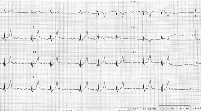
Chapter 68: Tricuspid Valve Dysplasia

Web Chapter 68


Tricuspid Valve Dysplasia


Tricuspid valve dysplasia is congenital malformation of the right atrioventricular valve apparatus and is thought to be caused by abnormal tissue undermining the right ventricle during embryogenesis. A variety of abnormalities are encountered, including thickened, shortened, or elongated leaflets; shortened or absent chordae tendineae; and abnormal papillary musculature. The major consequence of a malformed tricuspid valve is systolic regurgitation of blood into the right atrium, resulting in volume overload of the right side of the heart. This tricuspid valve regurgitation can lead to right-sided congestive heart failure and atrial arrhythmias.


Tricuspid valve stenosis occasionally may be a feature of the disease and is characterized by impaired diastolic opening as a result of thickened and fused leaflets. Tricuspid valve stenosis induces atrial hypertrophy and enlargement and also can lead to congestive signs and arrhythmias. Cyanosis and polycythemia may occur secondary to right-to-left shunting if a concurrent atrial septal defect or patent foramen ovale is present.


Ebstein’s anomaly is a related congenital defect in which the origins of the tricuspid leaflets are apically displaced into the right ventricle. It may or may not be associated with leaflet dysplasia. Ebstein’s anomaly has been documented only rarely in the veterinary literature (Chetboul et al, 2004; Takemura et al, 2003). Other congenital anomalies may be observed concurrently with tricuspid valve dysplasia, including mitral valve dysplasia, septal defects, pulmonic stenosis, and patent ductus arteriosus (Liu and Tilley, 1976). The foramen ovale may remain patent as a result of elevated right atrial pressure and dilatation.


Signalment


Tricuspid valve dysplasia is a relatively uncommon defect, accounting for approximately 3% to 7% of canine congenital cardiac defects in retrospective studies (Baumgartner and Glaus, 2003; Oliveira et al, 2011). Reports of tricuspid valve dysplasia have included a variety of large-breed dogs, such as Labrador retrievers, boxers, golden retrievers, Irish setters, Great Danes, and German shepherds (Andelfinger et al, 2003; Chetboul et al, 2004; Kornreich and Moïse, 1997; Liu and Tilley, 1976). The disease has been shown to be inherited in the Labrador retriever in an autosomal-dominant manner with incomplete penetrance and has been mapped to chromosome 9 in this breed (Andelfinger et al, 2003). However, a commercially available genetic test for breeding dogs is not available currently.


Tricuspid valve dysplasia is diagnosed less frequently in cats. It has been reported most commonly in the domestic shorthair cat but also has been noted in Chartreux and Siamese cats (Chetboul et al, 2004; Kornreich and Moïse, 1997; Liu and Tilley, 1976).


Diagnosis


Clinical Findings


Dogs and cats with tricuspid valve dysplasia may be clinically well with a heart murmur or may have right-sided congestive heart failure at presentation. Although most animals are evaluated at a young age because of the congenital nature of this defect, it is not uncommon to make the diagnosis in an older animal at the time that clinical signs develop.


The hallmark physical examination feature in animals with tricuspid dysplasia is a right apical systolic murmur. The intensity of the murmur can vary from very soft to very loud with a precordial thrill and depends on the amount of regurgitation and the velocity of regurgitant flow across the valve. Because of the latter factor, murmur intensity may not always reflect the severity of the disease. For example, a dog with a large tricuspid orifice resulting from severe malformation and volume overload of the right side of the heart may have nearly laminar regurgitant flow because of pressure equilibration between the right atrium and right ventricle. Some of these dogs have a very soft murmur. A soft diastolic murmur may be auscultated if tricuspid valve stenosis is present.


Dogs that are severely affected may have right-sided congestive heart failure with jugular venous distention or pulsation, hepatomegaly, and ascites at presentation. Femoral arterial pulse quality is expected to be normal unless the disease is very severe. Mucous membrane color is normal unless a coexistent right-to-left shunt is present. Animals with right-sided congestive heart failure often have poor body condition.


Electrocardiographic Findings


The most common electrocardiographic finding in animals with tricuspid valve dysplasia is a splintered QRS complex, with as many as two thirds of dogs and cats showing this abnormality (Kornreich and Moïse, 1997). Splintering describes an Rr′, RR′, rR′, or rr′ morphology of the QRS complex (Web Figure 68-1). The underlying mechanism for splintering is not known; however, possibilities include ventricular fibrosis resulting in altered conduction, right bundle branch conduction disturbances, and accessory pathway conduction.


< div class='tao-gold-member'>

Stay updated, free articles. Join our Telegram channel

Jul 18, 2016 | Posted by in PHARMACOLOGY, TOXICOLOGY & THERAPEUTICS | Comments Off on Chapter 68: Tricuspid Valve Dysplasia

Full access? Get Clinical Tree

Get Clinical Tree app for offline access