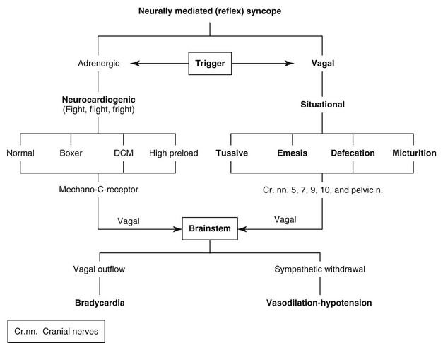
Chapter 67: Syncope

Web Chapter 67


Syncope


Syncope is a sudden but brief loss of consciousness from which recovery is spontaneous and complete. Presyncope (or near-syncope) refers to a brief, episodic rear limb or generalized weakness, ataxia, or altered level of consciousness. The common denominator is decreased or brief cessation of cerebral blood flow. Heart rhythm disturbances, either rapid or slow, are the most common causes of syncope. Rapid ventricular tachycardia (VT) and reflex bradycardias are most common; the latter also may be associated with reflex-mediated vasodilation. Arrhythmia-induced syncope in small dogs usually is the result of bradycardia (with or without vasodilation); in larger dogs it most often is related to VT. Syncope is less common in cats but can result from either tachycardia or bradycardias such as complete atrioventricular (AV block) (see Chapter 173).


Neurally Mediated Syncope


Terminology


Neurally mediated (reflex-mediated) syncope consists mostly of neurocardiogenic, vasovagal, and situational syncope. The terms neurocardiogenic and vasovagal syncope are considered synonymous by some, whereas others consider these separately. The authors prefer to use the term neurocardiogenic syncope when the reflex is believed to be triggered within the heart or pulmonary arteries. The precipitating event is an adrenergic (sympathetic) surge, and the resultant efferent activity most often resembles that of an exaggerated baroreceptor reflex characterized by bradycardia and vasodilation. Vasovagal syncope is common in humans and also can be triggered in the brain in response to emotional shock, phobias (such as blood phobia and needle phobia), instrumentation and pain.


Each neurally mediated episode of syncope can take one of three variations: marked slowing of the heart rate, inappropriate vasodilation, or a combination of the two events. When bradycardia dominates as the cause of hypotension and impaired cerebral perfusion, the term cardioinhibitory is appropriate. When vasodilation causes hypotension without an absolute bradycardia the term vasodepressor syncope is appropriate (with recognition that a relative bradycardia—or lack of reflex tachycardia—is common in these situations). Syncope associated with both bradycardia and vasodilation-hypotension comprises the mixed variant and represents the classic form of reflex-mediated syncope.


Situational syncope is neurally mediated and usually is triggered by a vagal surge rather than an adrenergic surge. Situational syncopes are those triggered by specific activities such as coughing (tussive), vomiting (emesis), defecation, or urination (micturition); these are named by the triggering event (e.g., tussive syncope). Situational syncope is most common in small dogs, and tussive syncope is the most common type. Situational syncope is uncommon in cats.


Carotid sinus syncope is a neurally mediated (reflex) event that is common in humans. It arises from mechanical pressure on the carotid sinus. Whether this syndrome occurs in dogs or cats is uncertain.


Pathophysiology


Neurally mediated syncope represents an exaggeration of cardiovascular reflexes that normally control the circulation. Syncope occurs when these reflexes intermittently become inappropriate in response to a trigger. The fact that some individuals develop reflex syncope commonly and sometimes with little provocation suggests that there is a disorder of autonomic control. The reflex is comprised of a trigger or afferent limb, a central nervous system integration system, and a response (efferent limb). Most often reflex syncope is classified by the afferent limb of the reflex arc.


When the afferent limb arises from adrenergic stimulation of receptors in the heart or pulmonary arteries the term neurocardiogenic syncope is appropriate. When the afferent limb arises centrally the term vasovagal is appropriate. Again, the response limb can be bradycardia with reduced cardiac output (cardioinhibitory), vasodilation (vasodepressor), or a mixed response. These responses can occur together but may persist to different degrees, as when transient bradycardia is associated with protracted hypotension owing to persistent vasodilation. At the center of the reflex is the cardiovascular control center in the medulla. The triggers and afferent pathways for these reflexes are incompletely understood. There appears to be a genetic predisposition in some humans. The same may be true in some dogs, especially the boxer.


Neurocardiogenic syncope is an adrenergically stimulated vagal reflex bradycardia. Vagal mechanoreceptors (C-fibers) initiate the reflex when stretched. These receptors are located in the ventricle, left atrium or left atrium–pulmonary vein junctions, and pulmonary arteries. Most episodes last only a few seconds to a minute. Respiratory arrest, white mucous membranes, and cyanosis occur during severe and protracted bradycardia. In some cases the owner believes that death has occurred.


The principal component of the bradycardia usually is sinus bradycardia proceeding to sinoatrial arrest followed by atrial, nodal, or ventricular escape beats. Sometimes periods of vagally induced AV block are found during these events. Episodes usually last for less than 1 minute, and as recovery begins the sinus rate increases but remains irregular and escape beats decrease in number. Following recovery a brief period of mild sinus tachycardia can occur. Because most episodes are brief, it may be difficult to document the bradycardia and even more difficult to document hypotension associated with peripheral vasodilation, although the mixed variant occasionally is documented. Mixed bradycardia-vasodilation is especially likely when a dog shows clinical signs of persistent hypotension in spite of a return to a slow-normal sinus rhythm.


Syncope associated with supraventricular tachycardia (SVT) and VT also may have a neurally mediated component. In this context SVT refers mostly to atrial tachycardia and AV reciprocating tachycardia. Atrial vasodepressor reflexes can result in significant vasodepression, causing a drop in blood pressure independent of the effect of the tachycardia.


Syncope or Seizure?


Signalment, known history of disease, situational triggers, and prodromal signs may provide clues to the origin of episodes or events. Collapse in a Doberman pinscher or boxer should be considered the result of rapid VT until proven otherwise, with recognition that some dogs of these breeds actually faint from bradycardia-vasodilation. Collapse episodes that are preceded by cough, gag, deglutition, emesis, micturition, or defecation most likely are situational syncope. Collapse in miniature schnauzers, West Highland white terriers, and American cocker spaniels often is the result of sick sinus syndrome. Collapse triggered by exertion-excitement in small dogs with advanced mitral valve degeneration-regurgitation or in apparently healthy dogs without evidence of heart disease often is a reflex (neurally mediated) event.


Although most often associated with flaccid collapse, syncope can be associated with extensor rigidity and spontaneous urination or defecation. Blanching of the oral mucosa always is present. Protracted bradycardia and respiratory arrest may result in cyanosis. Hypersalivation or facial fits rarely are associated with syncope. The exception is probably the cat, in which some facial movements are not uncommon and often resemble partial seizures. If, following an episode, the patient seeks water or food, then seizure should be suspected. Violent tonic-clonic motor activity seldom is the result of syncope, and seizure is more likely in such cases.


Causes of Syncope


Tachycardias


Supraventricular Tachyarrhythmias


Perhaps the most common supraventricular tachyarrhythmia is atrial fibrillation, although it is uncertain how often dogs faint from this rhythm disturbance. Atrial fibrillation usually is associated with advanced heart disease that has produced severe atrial dilation, generally affecting the left atrium. If the condition is untreated, the ventricular response rate in the hospital setting usually is 200 to 250 beats/min. New-onset atrial fibrillation can cause episodic weakness, collapse, or syncope. The loss of the atrial boost to left ventricular filling as well as rapid and particularly irregular diastolic filling durations, coupled with the underlying disorder, produce a marked drop in cardiac output.


Other causes of SVT include nodal tachycardia, AV reciprocating tachycardia, atrial flutter, and atrial tachycardia. Some of these SVTs can be associated with ventricular rates that exceed 300 beats/min and can be as high as 400 beats/min, especially with reciprocating tachycardias using an accessory pathway. The ventricular response rate to atrial flutter and some ectopic atrial tachycardias is quite variable because these often are associated with physiologic AV block that may control the ventricular rate response, sometimes to less than 200 beats/min. However, if AV nodal conduction is rapid, ventricular rates of 300 beats/min or higher can be seen. Rapid SVT is less likely to induce syncope because the rhythm is regular and the sequence of ventricular activation is normal, but collapse or loss of consciousness has been observed in some dogs. Many SVTs are not associated with overt structural heart disease, and structural changes may be due to the presence of tachycardia-induced cardiomyopathy. Of note is the SVT caused by AV reciprocating tachycardia (accessory pathway mediated) in the Labrador retriever.


Ventricular Tachycardia


One of the most common causes of syncope is rapid VT, especially in boxers with arrhythmic right ventricular cardiomyopathy (see Chapter 179) and Doberman pinschers with dilated cardiomyopathy (see Chapter 178). An inherited VT also occurs in certain families of young German shepherd dogs. Cardiac output and blood pressure can fall precipitously with rapid VT related to shortened diastole, AV dissociation, and aberrant sequence of ventricular activation. All dogs with dilated cardiomyopathy are at increased risk of VT, and sustained rates above 300 beats/min likely will degenerate into ventricular fibrillation and death. Boxers with arrhythmic right ventricular cardiomyopathy without myocardial failure often experience rapid VT and faint. However, as long as left ventricular function is normal, sudden death is unusual, even when VT is sustained. VT in animals with hypertrophic cardiomyopathy or subaortic stenosis can cause syncope and death at rates as slow as 180 beats/min.


In both VT and SVT, it has been shown in humans and experimental studies that the drop in blood pressure at the onset can be transient, and after several seconds the blood pressure can rise and syncope can resolve despite continued tachycardia. This stabilization results from reflex vasoconstriction and elevation of catecholamine levels.


Bradycardias


A number of heart rhythm disturbances can lead to bradycardia and syncope. These can include disturbances associated with neurally (reflex) mediated syncope as well as disorders of the impulse-forming and conduction systems of the heart.


Neurally Mediated Syncope


Neurally mediated (reflex) syncope probably is the most common bradycardia-associated syncope (Web Figure 67-1). Neurocardiogenic syncope is triggered by fight, flight, or fright scenarios and can occur in apparently healthy individuals or those predisposed by high preload, genetics (boxer), or dilated cardiomyopathy. The diagnosis often is presumptive.


< div class='tao-gold-member'>

Stay updated, free articles. Join our Telegram channel

Jul 18, 2016 | Posted by in PHARMACOLOGY, TOXICOLOGY & THERAPEUTICS | Comments Off on Chapter 67: Syncope

Full access? Get Clinical Tree

Get Clinical Tree app for offline access