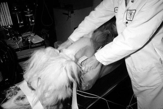
Chapter 60: Cardioversion

Web Chapter 60


Cardioversion


External electrical cardioversion refers to transthoracic delivery of a direct current (DC) shock for restoration of sinus rhythm in patients with ventricular or supraventricular tachyarrhythmias. The shock is synchronized to the R wave of the QRS complex to avoid induction of ventricular fibrillation. Synchronized energy delivery distinguishes cardioversion from defibrillation, which is unsynchronized. Until recently electrical cardioversion and defibrillation were done in humans and dogs using equipment that delivered a monophasic energy waveform (i.e., a high-energy electrical shock sent a current across the chest in a single direction). However, newer cardioversion/defibrillation devices are available that deliver biphasic energy waveforms. These biphasic cardioverters/defibrillators use a lower-energy self-reversing waveform that is safer and more effective than traditional monophasic shocks.


Biphasic electrical cardioversion has potential advantages over pharmacologic cardioversion in that a precisely regulated “dose” of electrical energy can restore sinus rhythm immediately and safely. Also, the distinction between supraventricular and ventricular tachycardia is less crucial with electrical cardioversion than with medical management. Finally, time-consuming titration of drugs with potential adverse effects is not an issue with DC cardioversion. However, a disadvantage of electrical cardioversion for emergency management of tachyarrhythmias is that the equipment and expertise may not be readily available. Moreover, when electrical cardioversion is done as an elective procedure, general anesthesia is required.


Electrical cardioversion appears to be most successful for termination of tachycardias resulting from impulse reentry, such as atrial fibrillation, atrial flutter, atrioventricular node reentry, atrioventricular reciprocating tachycardias involving an accessory conduction pathway, and most forms of ventricular tachycardia. The electrical shock is believed to terminate the tachycardia by simultaneously depolarizing all excitable myocardium and possibly by prolonging refractoriness; in other words, the shock interrupts reentrant circuits and establishes electrical homogeneity, thereby terminating reentry.


Indications and Contraindications for Direct Current Cardioversion


Electrical cardioversion should be considered for any tachycardia producing hypotension or congestive heart failure that does not respond promptly to medical management. The exception to this general rule is a digitalis-induced tachyarrhythmia.


DC cardioversion done as an elective procedure is useful for restoring sinus rhythm in dogs with sustained atrial fibrillation or atrial flutter. By improving hemodynamics (reducing ventricular filling pressures, augmenting cardiac output, decreasing ventricular rate, and providing atrioventricular synchrony), rhythm control generally is beneficial for dogs with congestive heart failure and secondary atrial fibrillation (Deedwania and Lardizabal, 2010; Sisson et al, 1995). Furthermore, restoration of sinus rhythm eliminates the need for pharmacologic control of heart rate using drugs that often have a negative inotropic effect, such as β-blockers and diltiazem (McNamara et al, 2003). In the absence of underlying heart disease, restoration of sinus rhythm in dogs with atrial fibrillation diminishes the risk of secondary thromboembolism and tachycardia-induced cardiomyopathy. In athletic dogs exercise tolerance also may improve. In dogs with atrial flutter, pharmacologic control of the ventricular rate frequently is difficult, and DC cardioversion often is the authors’ initial treatment of choice.


As discussed later in this chapter, the quality of life and heart failure status of dogs are improved greatly following successful cardioversion of atrial fibrillation to sinus rhythm. However, the likelihood of maintenance of normal sinus rhythm after successful cardioversion is affected adversely by the presence of structural heart disease as well as by chronicity of atrial fibrillation, and these factors should be taken into account by both the clinician and dog owner when considering cardioversion in a dog with congestive heart failure (see the later section on the duration of sinus rhythm after cardioversion of atrial fibrillation).


Electrical cardioversion is contraindicated in animals with cardiac glycoside toxicity, and there is an increased risk associated with electrical cardioversion in animals with sinus node dysfunction that do not have a pacemaker. Finally, cardioversion of atrial fibrillation in animals with intraatrial thrombus is contraindicated because resumption of an organized atrial contraction may cause embolization.


Cardioversion Procedure


Elective cardioversion is done under general anesthesia. Pretreatment with antiarrhythmic agents before performing electrical cardioversion in dogs with atrial fibrillation is recommended to help prevent recurrence of atrial fibrillation after cardioversion. Unless the dog’s condition is too critical to delay cardioversion, the authors treat for 2 weeks with amiodarone (12 to 15 mg/kg q24h PO) before the procedure. Although this antiarrhythmic agent causes pharmacologic restoration of sinus rhythm in some humans, the authors rarely observe this in dogs. Amiodarone is continued after cardioversion at a maintenance dosage of 5 to 7 mg/kg q24h PO. Preferably the cardioversion procedure is scheduled for early afternoon so that serum levels of electrolytes may be measured and electrolyte abnormalities corrected, if needed, immediately before induction of anesthesia. In addition the atria should be examined echocardiographically for thrombus in the morning on the day of the scheduled procedure. Successful cardioversion is more likely to be achieved if serum magnesium and potassium concentrations are in the middle to upper range of normal. The authors target a serum potassium concentration of 4.5 to 5.5 mEq/L and a serum magnesium concentration of more than 2 mEq/L. Digoxin should be withheld on the day of the procedure, but if digitalis toxicity is suspected, the cardioversion should be postponed until this problem has resolved.


Anesthetic premedication is recommended using an opioid (fentanyl 5 to 10 µg/kg, hydromorphone 0.05 to 0.2 mg/kg, oxymorphone 0.05 to 0.1 mg/kg, or methadone 0.5 to 1 mg/kg) either alone or in combination with a benzodiazepine (midazolam 0.1 to 0.3 mg/kg) administered SC or IM approximately 30 minutes before instrumentation and animal preparation. Before anesthetic induction the lateral thorax is clipped on each side from the axilla caudally and ventrally approximately 15 cm to facilitate placement and contact of the cardioversion paddles. Self-adhesive electrocardiographic (ECG) electrodes are attached to the skin of the extremities and connected to the ECG leads of the cardioversion device.


Anesthesia may be induced with fentanyl (5 to 20 µg/kg), etomidate (0.5 to 2 mg/kg), or a combination of these two drugs combined with a benzodiazepine (diazepam 0.2 to 0.5 mg/kg or midazolam 0.1 to 0.3 mg/kg). An anticholinergic agent administered at a conservative dose is indicated if the heart rate decreases enough to affect cardiac output and blood pressure adversely. Short-term maintenance of anesthesia for cardioversion may require additional doses of the induction drugs or supplementation with low doses of propofol (0.25 to 1 mg/kg).


The authors recommend that dogs be intubated and 100% oxygen administered using a standard small animal breathing circuit. Manual or mechanical ventilation of the animal may be necessary to avoid hypoventilation and respiratory acidosis during periods of deep sedation. Heart rate and rhythm, blood pressure, oxygen saturation, and end-tidal carbon dioxide concentration should be monitored during and immediately after cardioversion. Inhalation agents are used by some clinicians but generally are not administered in the authors’ practices unless the animal needs an additional diagnostic or surgical procedure following the cardioversion.


Although cardioversion may be accomplished by delivery of energy through either preplaced adhesive cardioversion pads or cardioversion paddles held against the thorax, the authors prefer paddles to achieve optimal positioning of electrodes and minimal transthoracic impedance. Immediately following induction of anesthesia, the dog is positioned in dorsal recumbency in a foam trough placed on a padded table. The forelimbs are secured cranially, and the rear limbs are secured caudally (Web Figure 60-1). Care must be taken to ensure that neither the dog nor the operator is in contact with any metallic surface.


< div class='tao-gold-member'>

Stay updated, free articles. Join our Telegram channel

Jul 18, 2016 | Posted by in PHARMACOLOGY, TOXICOLOGY & THERAPEUTICS | Comments Off on Chapter 60: Cardioversion

Full access? Get Clinical Tree

Get Clinical Tree app for offline access