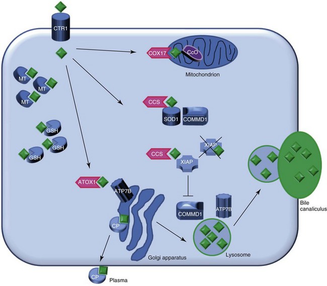
Chapter 48: Copper-Associated Hepatitis

Web Chapter 48


Copper-Associated Hepatitis


The essential trace element copper plays an important role in a variety of biologic processes, including antioxidant defense, mitochondrial respiration, iron metabolism, neurotransmitter synthesis, connective tissue formation, and pigmentation. Copper toxicity results from the presence of excess amounts of copper in the body. Free copper ions generate reactive oxygen species that can damage proteins, lipids, and DNA and also may have direct effects on cellular proteins. Therefore copper metabolism is regulated tightly.


The liver is the principal recipient of absorbed copper. In the liver, copper is stored and transferred to proteins for delivery to other tissues, and excess copper is eliminated via biliary excretion.


Copper-associated hepatitis can be the result of either an increased uptake of copper (i.e., high copper intake via diet or drinking water) or a decreased excretion of copper. A decreased excretion can be caused by a genetic defect (primary copper toxicosis), for example, COMMD1 deficiency in the Bedlington terrier or by cholestatic liver disease (secondary copper toxicosis).


Copper Metabolism


Copper metabolism is a complex process that is regulated on cellular and organ levels (Web Figure 48-1). Copper uptake takes place in the small intestine, where its absorption is mediated via copper transporter 1 (CTR1). The P-type ATPase ATP7A shuttles copper via the basal side of the enterocytes into the portal circulation. In the blood, small molecules such as histidine and serum proteins such as macroglobulin and albumin bind copper for transportation to the liver. CTR1 mediates hepatocellular uptake of copper. In the cytosol, copper is sequestered immediately by small molecules such as metallothionein and glutathione. Specialized copper chaperones shuttle copper to their destination proteins. The copper chaperone for superoxide dismutase (CCS) shuttles copper to superoxide dismutase (SOD1), a protein involved in oxidative stress defense and to XIAP, the X-linked inhibitor of apoptosis. Binding of copper to XIAP leads to an increased intracellular degradation of XIAP, which causes a lowering of the apoptotic threshold. COX17 is the chaperone for cytochrome C oxidase, which plays an important role in cellular respiration and in the mitochondria. ATOX1 delivers copper to ATP7B, which is necessary for copper incorporation into ceruloplasmin (CP) and facilitates copper excretion in bile. Mutations of ATP7B result in the human form of copper toxicosis: Wilson’s disease. COMMD1 can interact with ATP7B and works in conjunction with this protein in the process of copper excretion into bile. Other roles for COMMD1 in copper metabolism are under investigation and include a role in maturation and activation of SOD1 and the stabilization of ATP7A. COMMD1 function itself is in part regulated by XIAP.


Diagnosis of Copper-Associated Hepatitis


The best-described example of hereditary copper-associated hepatitis in the dog is autosomal-recessive Bedlington terrier copper toxicosis. In this dog breed, a deletion of the second exon of the COMMD1 gene results in a complete absence of the protein and a subsequent decrease of copper excretion into the bile leading to extreme accumulation of copper in the liver. The availability of a DNA test and implementation of this test in breeding strategies has led to an almost complete eradication of the disease. The DNA test can be based on the microsatellite marker C04107, which is located within the COMMD1 gene and where allele 2 is linked to the mutation. Since the discovery of the exact location of the mutation, direct detection of the deletion of exon 2 in genomic DNA is possible. There are incidental reports of affected Bedlington terriers that were heterozygous for the COMMD1 exon 2 deletion or had two copies of the wild type allele. In these dogs copper toxicosis may be explained by mutations in regulatory elements of COMMD1 or in a currently unidentified gene.


The increased incidence of copper-associated hepatitis in certain other breeds is a strong indication for a hereditary background. Pedigree studies executed in the Labrador retriever, Doberman pinscher, West Highland white terrier, Skye terrier, and Dalmatian confirmed a hereditary background. In these breeds a complex form of copper-associated hepatitis is expected, where the susceptibility for copper is determined genetically, and the expression of the disease phenotype may rely on environmental factors such as dietary copper intake. In the Doberman pinscher there is increasing evidence for involvement of deregulation of autoimmune processes in the cause of Doberman hepatitis. No DNA test exists for breeds other than the Bedlington terrier.


Signalment


Besides the increased incidence of copper-associated hepatitis in the aforementioned dogs, other dogs also can be affected. In a recent review of dogs diagnosed with primary hepatitis, roughly one third of cases were diagnosed with copper-associated acute or chronic hepatitis based on histologic evaluation of rubeanic acid–stained slides of liver tissue. Breeds diagnosed with primary copper-associated hepatitis include German shepherd, Anatolian shepherd, Keeshond, boxer, Airedale terrier, Norfolk terrier, beagle, American cocker spaniel, cavalier King Charles spaniels, as well as mixed-breed dogs. No prospective studies of the family members of these dogs were performed to establish a heritable background; however, the absence of evidence for extremely high dietary intake of copper in these dogs and the centrolobular localization of copper indicate a primary copper storage disease.


Sex distribution may be different between breeds. In the Labrador retriever and Doberman pinscher a strong female predisposition for copper-associated hepatitis is present, whereas in Bedlington terriers, West Highland white terriers, Skye terriers, and Dalmatians an equal distribution between sexes exists.


The age at which dogs are presented may vary. Hepatic copper accumulation often starts before the age of 1 year, but the appearance of clinical signs may be delayed. Dogs with inherited copper storage disorders may be subject to a prolonged initial delay period between copper accumulation and the development of histologic evidence of inflammation and a second delay between first histologic evidence of inflammation and recognition of clinical symptoms of disease. This last delay may be due to the fact that clinical signs usually appear when a large part of the liver capacity is affected often in the end stage of disease. The speed of progression of disease may depend on environmental factors: for example, dietary intake and gestation and lactation in bitches. In general, first clinical presentation can range from the age of 2 years to the age of approximately 12 years.

< div class='tao-gold-member'>

Stay updated, free articles. Join our Telegram channel

Jul 18, 2016 | Posted by in PHARMACOLOGY, TOXICOLOGY & THERAPEUTICS | Comments Off on Chapter 48: Copper-Associated Hepatitis

Full access? Get Clinical Tree

Get Clinical Tree app for offline access