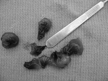
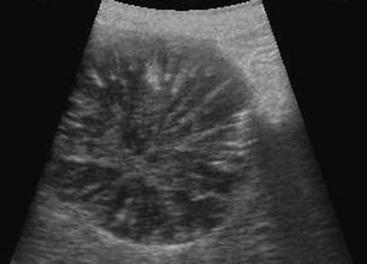
Web Chapter 46 A biliary mucocele is an accumulation of the mucus component of bile within the gallbladder (GB). Grossly it appears as shiny green-black gelatinous material with lamellar striations along fracture lines (Web Figure 46-1). Functional or structural obstruction of the cystic or common bile duct is the usual cause in humans and was proposed initially as a cause in the dog. However, studies do not support this theory. Primary obstruction of the cystic or common bile ducts rarely is found on diagnostic tests or at the time of surgery, and histologically there is typically evidence of hyperplasia of the mucus-secreting glands of the GB mucosa. Current causative theories are multifactorial. Impaired gallbladder motility and cholesterol and lipid metabolism appear to be primary factors in disease development. Delayed GB motility allows the accumulation of concentrated bile salts, which stimulate mucus production from the endothelial lining. Diabetes mellitus, hypothyroidism, and exogenous steroid administration (androgenic and corticosteroids) have been associated with delayed GB emptying. A correlation between cholesterol and mucin content of bile also has been established, indicating that hypercholesterolemia and poor lipid metabolism could be contributors. One study has shown that dogs with hyperadrenocorticism are 29 times more likely to develop a biliary mucocele than dogs without hyperadrenocorticism, and a trend for association with hypothyroidism also was suggested (Mesich et al, 2009). A predisposition for endocrinopathies and abnormalities in lipid metabolism may explain the breed predisposition associated with biliary mucoceles (Shetland sheepdogs, cocker spaniels, miniature schnauzers, and dachshunds). More recently researchers have found an association of a mutation in ABCB4 transporter gene in Shetland sheepdogs and other dogs having biliary mucoceles (Mealey et al, 2010). This gene functions in transporting phosphatidylcholine across the canalicular membrane. Abnormal bile composition changes may be in part responsible for damage to the biliary endothelial lining and subsequent gallbladder mucinous hyperplasia. Web Figure 46-1 A biliary mucocele with green-black gelatinous mucus material and lamellar striations along fracture lines. Patients typically present as middle-aged to older, small- to medium-breed dogs. A breed predisposition for Shetland sheepdogs and an overrepresentation of dachshunds, miniature schnauzers, and cocker spaniels have been reported (Aguirre et al, 2007). No sex predilection is evident. The most common laboratory abnormalities include increases in serum liver enzyme activity (alkaline phosphatase, alanine aminotransferase, alanine aspartate aminotransferase, γ-glutamyltransferase, increased total bilirubin) and elevated white blood cell counts. Abdominal radiographs have limited usefulness in the diagnosis of a biliary mucocele but may be critical in ruling out other causes of the historical or physical findings such as gastrointestinal obstruction, abdominal mass, or other causes of vomiting and lethargy. Although uncommon, choleliths may be visible on abdominal radiographs, and these occasionally may lead to biliary tract obstruction. Abdominal ultrasound is the preferred diagnostic test for GB mucoceles, which have distinct characteristics on ultrasonographic images. The tenacious mucin along the GB wall is visualized as a hypoechoic rim. The central contents are hyperechoic and non–gravity dependent with ballottement or repositioning of the patient. Radiating striations extend from the GB wall centrally as the mucocele matures, creating the described “kiwi fruit” appearance specific to mucoceles (Web Figure 46-2). Ultrasound-guided cholecystocentesis is contraindicated for biliary mucoceles. Ultrasonography also may detect varying degrees of extrahepatic biliary obstruction caused by extension of the mucinous bile into the cystic, common, and hepatic ducts. Although the ultrasound examination can be sensitive for the diagnosis of biliary mucoceles, it does not always correspond as favorably to the severity of disease assessed at surgery, in particular the presence of pressure necrosis of the GB wall or cystic duct that can lead to eventual rupture. Evidence of necrosis or rupture include hyperechoic fat and/or a hypoechoic halo of fluid surrounding the GB. Web Figure 46-2 Ultrasound view of a gallbladder mucocele demonstrating the characteristic striated or stellate (kiwi-shaped) pattern within the lumen of the gallbladder.
Canine Biliary Mucocele
Pathogenesis

Diagnostics

![]()
Stay updated, free articles. Join our Telegram channel

Full access? Get Clinical Tree


