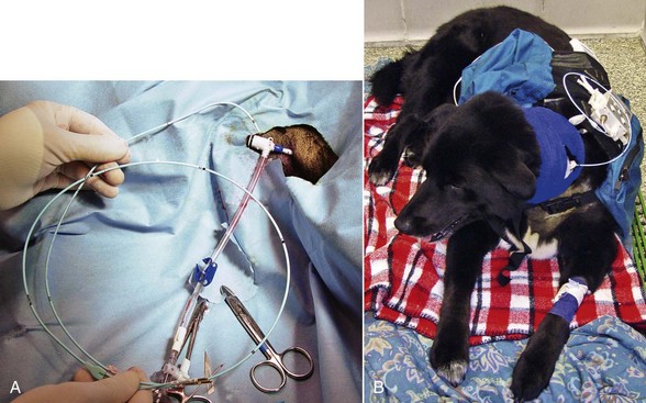
Chapter 4: Pacing in the ICU Setting

Web Chapter 4


Pacing in the ICU Setting


Transvenous pacing (TVP) and transcutaneous external pacing (TCP) are temporary pacing techniques that can be lifesaving in the emergency setting. Both procedures are performed in acute situations to treat critically ill patients in need of immediate control of the heart rhythm.


Hemodynamically significant bradyarrhythmias may be encountered in the critical care environment related to a variety of causes. The majority of veterinary patients treated by temporary cardiac pacing are symptomatic for the rhythm disturbance and awaiting implantation of a permanent cardiac pacemaker. Less frequently, prophylactic placement of a temporary pacemaker is used to prevent hemodynamic instability due to drug toxicity or in the perioperative setting as with cardiac surgery or elective surgery in dogs with medically treated sick sinus syndrome. Certain tachyarrhythmias also can be treated with temporary cardiac pacing if they are unresponsive to medical intervention, but this is much less common.


The choice between TVP and TCP should be made according to equipment availability and personal proficiency in the use of pacing devices. TVP requires more specialized training and additional equipment such as pacing leads, an external pulse generator, and ideally fluoroscopy. Placement of TVP leads without fluoroscopic guidance is possible by using simultaneous electrocardiography (ECG) monitoring (with or without ultrasound guidance). However, in most cases fluoroscopic guidance is quicker and results in a more secure lead position for temporary cardiac pacing. In contrast, TCP is noninvasive, is relatively easy to perform, does not include concerns relative to radiation safety, and requires very little preparation time. TCP is especially useful in patients that have experienced cardiopulmonary arrest, in those already under anesthesia, and in situations in which severe cardiovascular collapse requires immediate intervention. However, TCP is not ideal if temporary pacing is needed for more than a few hours because the discomfort and pain associated with the procedure mandate general anesthesia during cardiac pacing.


If equipment for either TCP or TVP is unavailable, clinicians can attempt to increase the patient’s heart rate with drugs such as atropine, theophylline, terbutaline, or isoproterenol. However, these medications may have proarrhythmic effects that might cause serious ventricular arrhythmias, and most importantly medical treatment is often unsuccessful. Catecholamines also can exert unacceptable peripheral vascular effect as with isoproterenol-induced vasodilation. In most cases of severe symptomatic bradycardia, immediate referral to a hospital with the capability to perform cardiac pacing is medically indicated.


Indications for Temporary Pacing


Temporary pacing is probably most commonly performed immediately prior to implantation of a permanent cardiac pacemaker. In this situation, temporary pacing can prevent marked bradycardia or asystole associated with induction of anesthesia, and this may be the most common use of temporary pacing. In the case of TCP, the cutaneous pacing electrodes are placed but the system is not activated until unconsciousness or general anesthesia has been induced. Additionally, several emergency and critical care situations are also indications for temporary cardiac pacing. These cases fit into one of three categories: bradyarrhythmia with signs of low cardiac output, bradyarrhythmia with congestive heart failure (CHF), or bradycardia encountered during cardiopulmonary arrest.


Bradyarrhythmia with Low Cardiac Output


Although bradycardias are commonly encountered in clinical practice, not all bradyarrhythmias require immediate drug therapy or cardiac pacing. Stable cases with isolated weakness or infrequent, milder forms of syncope may be able to wait for a short time until elective pacemaker implantation is feasible, especially for dogs affected with sick sinus syndrome. Emergency cardiac pacing, however, is indicated in symptomatic bradyarrhythmias that result in profound weakness, progressive azotemia, frequent or severe syncope, or progressive elevations of blood lactate. Animals with bradycardia and moderate-to-marked elevations of blood lactate often require urgent temporary cardiac pacing.


Arrhythmias that commonly lead to low cardiac output severe enough to require temporary cardiac pacing include high-grade atrioventricular (AV) block or sinus arrest and very slow ventricular escape rhythms (< 35 to 40 per minute). Animals with long pauses (e.g., > 8 to 12 seconds) in cardiac rhythm that result in weakness, collapse, or syncope may require emergent cardiac pacing. The simultaneous presence of malignant ventricular arrhythmias and complete AV block represents another situation in which pacing may either alleviate the tachyarrhythmia or permit safe use of antiarrhythmic drugs. Bradyarrhythmias that occur during anesthesia and those encountered with certain toxicities, especially overdose of calcium channel blockers, often respond well to temporary cardiac pacing.


Dogs with sinus arrest may experience multiple syncopal episodes, but they are less likely to experience sudden death than dogs with third-degree AV block. In addition, dogs with sinus arrest are more likely to respond favorably to pharmacologic management (anticholinergics or sympathomimetics), especially in the emergency or perioperative setting, which can often preclude or delay the use of cardiac pacing. In contrast, a permanent pacemaker is recommended for dogs with third-degree AV block and signs of syncope or low cardiac output. Attempts to medically treat this arrhythmia should be limited to the hospital. Atropine rarely works, although one dose can be tried. Catecholamines are rarely effective in treating AV block, are proarrhythmic in patients with bradycardia, and most importantly delay potentially life-saving pacing therapy.


Once the temporary pacing device has been placed and ventricular capture is established, the rate on the pulse generator is usually set between 70 and 90 pulses per minute for dogs. This rate is usually adequate to prevent syncope and meet hemodynamic demands at rest.


Bradyarrhythmia with Congestive Heart Failure


In animals with simultaneous bradycardia and evidence of congestive cardiac failure, temporary pacing can be a successful adjunct to standard heart failure treatments. Although most patients with sinus node dysfunction or AV block do not present with CHF, dogs with profound bradycardia may develop pulmonary edema, pleural effusion, or ascites. Some of these dogs have concurrent structural disease; for example, most dogs with sick sinus syndrome tend to have some degree of chronic degenerative valve disease. Some benefit significantly from pacing along with medical therapy of CHF, and in those without structural heart disease such as dilated cardiomyopathy or severe mitral regurgitation, pacing may prevent redevelopment of CHF. An exception may be those dogs with atrial standstill due to atrial muscular dystrophy in which pacing may prevent syncope but cannot reverse the severe underlying myocardial disease.


Because the long-term prognosis after permanent cardiac pacing for dogs with bradycardia and concurrent CHF may not be quite as good as for dogs with isolated bradycardia, assessment of overall cardiac status with echocardiography is important to determine the extent of concurrent structural cardiac disease prior to permanent cardiac pacing. In the dog with bradycardia and concurrent CHF, temporary pacing can be performed for 1 to 4 days until signs of CHF have improved, the benefit of pacing is more clear, and the dog becomes a better candidate for general anesthesia. The external pulse generator is usually set at a rate between 120 and 140 pulses per minute until signs of CHF begin to resolve.


Cardiopulmonary Arrest


During cardiopulmonary arrest (see Chapter 5) early identification of bradycardia and rapid initiation of a temporary pacing may supplement standard cardiopulmonary resuscitation (CPR) and may be beneficial for some patients. Arrhythmias that may benefit from cardiac pacing include severe sinus bradycardia, advanced AV block, slow ventricular escape rhythms, and asystole. Cardiac pacing is usually not effective in animals with pulseless electrical activity (e.g., electromechanical dissociation) or for ventricular fibrillation. During CPR, pacing is usually attempted when atropine and repeated doses of epinephrine administration fail to increase ventricular firing to a rate that is capable of providing adequate perfusion for the patient.


For pacing to be successful, ventricular capture must occur. Both ventricular capture and myocardial contractions from pacing are negatively affected by prolonged CPR as underlying myocardial ischemia, hypoxia, acid-base disturbances, and electrolyte abnormalities become more profound. In such cases, electrical capture may still occur at high current outputs, but the result may be pulseless electrical activity without effective ventricular contraction.


Placement of the transvenous catheter during cardiopulmonary arrest is usually attempted without fluoroscopy, and with the motion artifact that occurs on the ECG during CPR, the procedure can be exceedingly difficult. However, if open-chest CPR has been performed, the heart can often be paced directly by placing the pacing lead on the epicardial surface of the heart. The heart can be paced with this technique until the patient’s cardiac rhythm is stable or jugular vein access has been secured for transvenous placement of the lead. During CPR, TCP may be preferred if a defibrillator with transcutaneous pacing capability is available. The pacing rate may be dictated by other clinical parameters, but initially a pacing rate of 100 to 120 beats per minute or greater is recommended for cardiac pacing during CPR.


Temporary Transvenous Pacing


Temporary TVP is minimally invasive and can be performed within a short time by trained personnel. However, the approach requires significant knowledge of the cardiovascular system and catheter manipulation, and TVP can be associated with small but significant risk. The success rate and incidence of complications are highly influenced by the experience of the clinician. Therefore emergency and critical care veterinarians should understand the indications, equipment, techniques, and complications associated with the procedure.


Transvenous Pacing Technique


Insertion techniques can include the use of fluoroscopic imaging, intracavitary ECG monitoring, or blind advancement in the jugular vein with surface ECG monitoring. Fluoroscopy is usually needed for placement of stiff, nonfloating catheters, while in many cases flow-directed catheters can be advanced with some combination of ECG, echocardiographic, or fluoroscopic guidance. Balloon catheters may decrease procedure time and improve lead positioning. The ECG-guided technique is challenging, and prior experience with transvenous pacemaker implantation using fluoroscopy is advantageous before attempting ECG guidance alone.


Patient Preparation and Sedation


A temporary pacemaker can usually be placed with the patient under light sedation and local anesthesia. Opiates, combined with a benzodiazepine, produce excellent mild sedation (i.e., oxymorphone, 0.05 to 0.1 mg/kg IV, or buprenorphine, 0.005 to 0.01 mg/kg IV in combination with diazepam, 0.1 to 0.2 mg/kg IV). General anesthesia may be needed in selected cases, but this is usually avoided due to concerns that induction of anesthesia could lead to severe bradycardia or asystole. Perioperative antibiotics should be administered to the patient if it is not on antibiotics at the time of the procedure.


The right or left jugular vein, the femoral vein, or the saphenous veins can be used for placement of the pacing lead, depending on patient size and vascular access. In some dogs, a 4 or 5 French pacing wire may be successfully advanced up the lateral saphenous vein. Jugular vein access is easier, and the lead is less likely to migrate or dislodge. The right jugular vein is the more direct route to the right atrium and ventricle and avoids any issues related to a persistent left cranial vena cava, but the left jugular vein or femoral vein should be used for temporary lead wire placement if a future permanent transvenous pacemaker implantation is anticipated (as the right jugular vein is generally chosen for permanent pacing). The skin over the target vein is first clipped, quickly cleaned, and infiltrated with 2% lidocaine, after which the skin is aseptically prepared. The vessel can be accessed via a cut-down technique or by vascular puncture using a catheter sheath introducer system (5 to 8.5 French percutaneous introducer) and a modified Seldinger approach (Web Figure 4-1, A). Once vascular access is secured, the pacing lead is passed into the vessel and toward the heart. Some operators prefer to insert the pacing catheter into a telescoping plastic sleeve that can later be advanced to cover the catheter and interlocked with the vascular access sheath; this allows for repositioning with less risk of contamination. Continuous electrocardiographic monitoring is performed during lead insertion to identify correct pacing lead placement, ventricular capture, or ventricular ectopy that might require repositioning of the catheter. Difficulties in lead advancement can be encountered with variations in vascular anatomy (e.g., persistent left cranial vena cava).


< div class='tao-gold-member'>

Stay updated, free articles. Join our Telegram channel

Jul 18, 2016 | Posted by in PHARMACOLOGY, TOXICOLOGY & THERAPEUTICS | Comments Off on Chapter 4: Pacing in the ICU Setting

Full access? Get Clinical Tree

Get Clinical Tree app for offline access