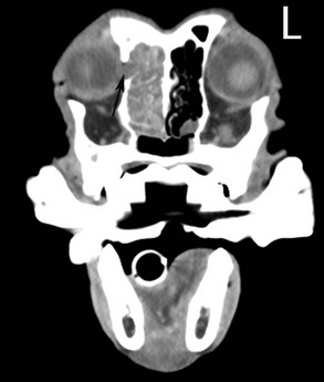
Web Chapter 29 1. Straight lateral to medial view 2. Open-mouth ventrodorsal maxillary view, angled 10 degrees rostral-caudal 3. Intraoral dorsoventral maxillary view However, cross-sectional imaging is preferred. Computed tomography (CT) is the most commonly used imaging modality for evaluating dogs and cats with nasal disease, but magnetic resonance imaging (MRI) also is used. Both imaging modalities have advantages and disadvantages in the imaging of nasal disease in cats and dogs. Bone destruction is easier to identify on CT (Web Figure 29-1), and soft tissue changes are delineated better on MRI. Generally MRI studies are more expensive, and imaging time is longer than CT studies. Because computer-based radiation therapy (RT) plans are generated commonly using CT images, CT may be the preferred imaging modality if RT is contemplated. Web Figure 29-1 Postcontrast computed tomographic (CT) axial slice at the level of the eyes of an 8-year-old field spaniel dog with a right-sided nasal adenocarcinoma. Note the soft tissue attenuating material (tumor) in the right nasal cavity. Tumor destruction of the right maxilla (black arrow) with tumor extension into the right retrobulbar space is present.
Nasal Tumors
Staging and Diagnosis
Imaging
< div class='tao-gold-member'>
![]()
Stay updated, free articles. Join our Telegram channel

Full access? Get Clinical Tree


