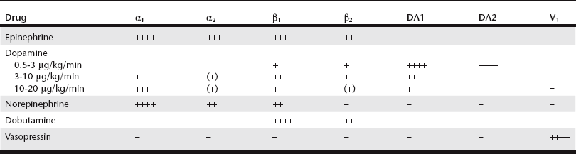
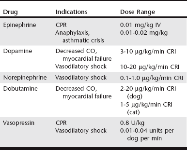
Catecholamines in the Critical Care Patient

Chapter 3


Catecholamines in the Critical Care Patient


Catecholamines are the primary effectors of the sympathetic nervous system (SNS) and play a major role in cardiovascular homeostasis in normal and pathophysiologic stress. Physiologic sympathetic outflow, which is maintained through a basal level of stimulation from the vasomotor center and adrenal secretion of catecholamines, sustains appropriate heart rate, vascular tone, and blood pressure. Endogenous catecholamines include norepinephrine (NE) and dopamine (DA), both primarily released by sympathetic nerve fibers, and epinephrine (EPI), primarily released by the adrenal gland. A number of stimuli, including pain, fear, hypoxemia, anemia, and hypotension, can increase the release of these sympathetic mediators and subsequent activation of adrenergic receptors. The resulting “fight or flight” response is characterized by tachycardia, increased cardiac output, and elevation of blood pressure.


The actions of catecholamines are largely determined by the location and type of receptor activated. These receptors are classified as presynaptic or postsynaptic. Presynaptic receptors have a regulatory effect on the amount of NE released from the nerve terminal, increasing or decreasing it depending on the receptor type. Postsynaptic receptors are subdivided into synaptic/junctional (having the most significant role in tissue activation) and extrajunctional (only stimulated with massive catecholamine release). A number of different receptor types (and subtypes) have been elucidated and include α-adrenergic (α1, α2), β-adrenergic (β1-3), and dopaminergic (DA1, DA2). Each receptor type has a relative distribution within certain tissues, a varying affinity for the adrenergic neurotransmitters, and specific physiologic effects when activated.


Catecholamines exert their effects by binding with a receptor and then triggering intracellular signaling pathways. Although these effects are complicated and modulated by vagal input, circulating hormones, and locally produced mediators, some general points deserve emphasis. Vascular postsynaptic α1 and α2 receptors increase intracellular calcium levels, leading to vasoconstriction. Presynaptic α2 receptors diminish the release of NE and promote down-regulation of sympathetic outflow, with resultant vasodilation and diminished cardiac output. β Receptors modify cyclic adenosine monophosphate (cAMP) levels, which alter cytosolic calcium levels depending on the tissue location. Postsynaptic β1 receptors, which are largely located on myocardial cells, increase intracellular (cytosolic) calcium, leading to increased contractility (inotropy) along with enhanced reuptake of calcium in diastole that speeds myocardial relaxation (lusitropy). In specialized tissues such as the sinoatrial (SA) and atrioventricular (AV) nodes the postsynaptic β1 receptors both enhance calcium entry and stimulate funny current to increase heart rate (chronotropy) and speed of conduction across the SA and AV nodes. Myocardial β2 activation has mostly similar effects, although these receptors are extrajunctional and require significant stimulation. β2 receptors located in bronchial and vascular tissues promote vascular smooth muscle relaxation, which results in bronchodilation and vasodilation. For tissues with multiple types of adrenergic receptors, the net effect of activation depends on receptor prevalence and affinity for catecholamine stimulation.


Specific Catecholamines


Given the varied effects and tissue beds that are subject to sympathetic stimulation, there is significant potential for therapeutic manipulation through administration of exogenous catecholamines. In critical care patients these medications are primarily used to augment catecholamine effects on the cardiovascular system, especially when the endogenous response might be insufficient or become exhausted. Determining which catecholamine to use is largely based on its relative receptor activities (Table 3-1), the clinical scenario in which intervention is needed (Table 3-2), and the potential for adverse effects.


Epinephrine


EPI is nonselective in its receptor activation and has effects on all α and β receptors. The α effects primarily cause vasoconstriction, although the net impact on peripheral resistance is partially offset by β2-induced vasodilation. The β effects promote an increase in cardiac output as well as bronchodilation. Given this combination of activities, EPI is primarily used in the treatment of patients with cardiopulmonary arrest, anaphylaxis, or severe asthmatic crisis. Potential adverse effects include excessive vasoconstriction, especially to the splanchnic circulation; increased myocardial oxygen demand; arrhythmias; tachycardia; hyperglycemia; and hypokalemia.


The primary beneficial effect of EPI in cardiopulmonary resuscitation (CPR) is α-induced vasoconstriction (and resultant increase in diastolic pressure) rather than any direct cardiac effects from β stimulation. Coronary circulation is an essential component of successful resuscitation, and diastolic pressure is a major determinant for coronary perfusion pressure. In addition, EPI has beneficial effects on cerebral perfusion and oxygen delivery. Despite these potential benefits, available evidence shows no clear survival benefit with administration of EPI in CPR (Neumar et al, 2010). However, EPI remains the drug of choice in CPR because it has been shown to improve rates of return of spontaneous circulation (ROSC).


Ideal dosing has been a subject of some debate. Although it has increased α1 stimulation, “high-dose” EPI (0.1 mg/kg) has not proved any more effective in achieving ROSC than “low-dose” EPI (0.01 mg/kg) in most clinical circumstances of arrest. Also, with “low-dose” EPI some of the adverse effects of EPI are avoided in the postresuscitation period, especially if repeated doses are needed, and this “low-dose” approach currently is recommended in CPR (Neumar et al, 2010). One must be mindful of the concentration of epinephrine used (1 : 1000 vs 1 : 10,000). For “low-dose” EPI, standard dosing with 1 : 10,000 is 0.1 ml/kg (or 1 ml/10 kg). If only 1 : 1000 is available, it should be diluted 1 : 10 in sterile water and then administered as above. This can be repeated every 3 to 5 minutes (after each one to two cycles of compressions) until ROSC or cessation of CPR. When administered via the endotracheal route if intravenous (IV) access is unavailable, doses administered during CPR should be doubled.


Another potential application of EPI is in the treatment of anaphylaxis/anaphylactic shock. The combination of vasoconstriction and bronchodilation makes EPI, along with IV fluids and oxygen therapy, ideally suited to help reverse the major life-threatening aspects of anaphylaxis. In addition, EPI serves to decrease production and release of mediators involved in the pathogenesis of anaphylaxis. Although there is some evidence of improved outcomes with a constant rate infusion (CRI) of EPI (Mink et al, 2004), bolus dosing of 0.01 to 0.02 mg/kg IV (potentially repeated after 15 to 20 minutes) is still currently recommended. EPI also may be beneficial as a bronchodilator for cats in asthmatic crisis, dosed at 0.01 to 0.02 mg/kg IV or subcutaneously (SC).

< div class='tao-gold-member'>

Only gold members can continue reading. Log In or Register to continue

Stay updated, free articles. Join our Telegram channel

Jul 18, 2016 | Posted by in PHARMACOLOGY, TOXICOLOGY & THERAPEUTICS | Comments Off on Catecholamines in the Critical Care Patient

Full access? Get Clinical Tree

Get Clinical Tree app for offline access