Carbon Monoxide

Chapter 87 Carbon Monoxide


PATHOPHYSIOLOGY


Hypoxic Toxicity


The hypoxic mechanism of carbon monoxide toxicity involves the interaction of carbon monoxide with hemoglobin in the blood: carbon monoxide displaces oxygen from hemoglobin and causes an allosteric hindrance of oxygen release from hemoglobin to the tissues.4,16 The resultant tissue hypoxia results in cellular shock, CNS depression, and cardiovascular compromise.


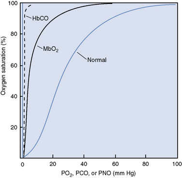
Carbon monoxide competes with oxygen for hemoglobin binding sites with 200 to 250 times the affinity.1,16 Carbon monoxide binds two of the four available heme groups in each molecule of hemoglobin, causing a decrease in the oxygen carrying capacity of 50% (Figure 87-1) and shifting the oxyhemoglobin dissociation curve down.16,17 Thus very low levels of carbon monoxide in the blood result in markedly reduced oxygen carrying capacity despite a normal hemoglobin concentration and normal partial pressure of oxygen.


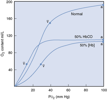
The dissociation of oxygen from hemoglobin is also affected by carbon monoxide, which binds tightly to hemoglobin, markedly disturbing the chemical equilibrium of the molecule.16,17 The change in equilibrium of the COHb results in an interference with both the association and dissociation of oxygen.16 Therefore oxygen bound to hemoglobin, which is also bound to carbon monoxide is not easily released to the tissues, and the oxyhemoglobin dissociation curve is shifted to the left (Figure 87-2).16,17 Decreased release of oxygen to the tissues exacerbates the cellular hypoxia caused by decreased oxygen content and subsequent delivery.


Cellular Toxicity


Hypoxia alone, however, does not explain the variation in individual response to a given level of COHb and the delayed neurologic syndrome associated with carbon monoxide toxicity. Studies have demonstrated cerebral hyperemia, tachycardia, and tachypnea during carbon monoxide toxicity, representing a compensatory mechanism to maintain oxygenation in the face of decreased oxygen carrying capacity.1,7,18 Therefore the brain does not commonly suffer from hypoxia during carbon monoxide toxicity unless there is accompanying cardiovascular dysfunction or severely elevated levels of COHb (>70%), overwhelming the adaptive response.7,18 A study was performed comparing the clinical signs of dogs subjected to inhaled carbon monoxide with dogs bled to a hematocrit of approximately 25% and subsequent volume replacement with crystalloid and colloid fluids or COHb-containing red blood cells.19 The dogs subjected to inhaled carbon monoxide achieved COHb levels of 54% to 90% and died. The dogs bled to an anemic state and given COHb-containing red blood cells to achieve a COHb level of 60% survived indefinitely with an outcome similar to that of dogs who were bled and received volume replacement with crystalloid and colloid solutions.19 This study confirmed the suspicion that a mechanism other than hypoxia from high levels of COHb results in toxicity. This mechanism is thought to be the consequence of cellular toxicity.2,4,7,19 Cellular toxicity associated with carbon monoxide poisoning appears to involve both direct and indirect mechanisms.


Indirect Mechanisms


There is a growing body of evidence demonstrating indirect mechanisms through which carbon monoxide toxicity results in changes that lead to cellular toxicity, particularly within the brain. These changes include sequestration of leukocytes,7 increased nitric oxide (NO) production,7,21,22 reperfusion injury,1,7 lipid peroxidation,1,7,23 and direct neurotoxicity from carbon monoxide activity as the a neurotransmitter.6,7


It has been demonstrated experimentally that carbon monoxide activates polymorphonuclear leukocytes (PMNLs), resulting in diapedesis and leukoencephalopathy.7 The activated neutrophils can contribute to tissue damage by producing reactive oxygen species, releasing proteolytic enzymes, and obstructing capillaries as the PMNLs accumulate.24 NO levels increase during exposure to high levels of carbon monoxide as a result of increased NO release from platelets21 and production in neuronal tissue.7,22 NO may have both a protective and damaging role in carbon monoxide toxicity. NO decreases PMNL adhesion to endothelium, and a transient rise in nitric oxide may protect brain tissue initially, although falling levels may be responsible for the delayed neurologic sequelae seen in some cases.21 The combination of NO and superoxide (one of the reactive oxygen species produced in activated PMNLs and other cells during reperfusion), however, results in peroxynitrite production, a key player in lipid peroxidation.24 Lipid peroxidation is a process characterized by conversion of membrane lipids to reactive species and propagation of oxidative damage, culminating in severely damaged or destroyed cell membranes.24 Thus elevated carbon monoxide levels set up a chain reaction resulting in direct tissue damage, reperfusion injury, lipid peroxidation, cellular dysfunction, and ultimately cell death within the brain.7,23


The role of endogenous carbon monoxide as a neurotransmitter and the potential implications of exogenous carbon monoxide overstimulating certain neural functions are being explored.6,7 An altered ratio of carbon monoxide and NO may exaggerate neurologic malfunction.7

Stay updated, free articles. Join our Telegram channel

Sep 10, 2016 | Posted by in SMALL ANIMAL | Comments Off on Carbon Monoxide

Full access? Get Clinical Tree

Get Clinical Tree app for offline access