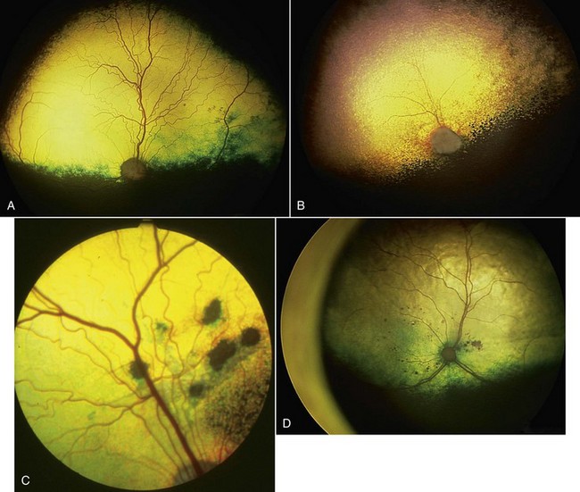
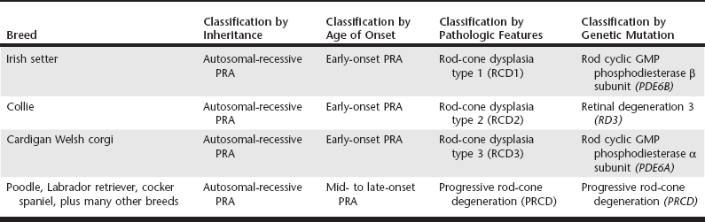
Chapter 254 Retinopathies in dogs may be identified because of a loss of vision or an altered external appearance of the eyes. Many cases are detected only during a careful ophthalmoscopic examination (Figure 254-1) as part of a general physical examination or at a screening clinic (e.g., a Canine Eye Registration Foundation examination). This chapter considers hereditary and acquired diseases of the canine retina. Figure 254-1 A, Wide-angle view of a fundus of a normal adult dog. B, Wide-angle view of a fundus of a Cardigan Welsh corgi with progressive retinal atrophy. The tapetal fundus is hyperreflective due to retinal thinning and the retinal vasculature is attenuated. C, Fundus of a springer spaniel with geographic retinal dysplasia lesions. The lesions have an altered tapetal color and the larger ones have a pigmented center. D, Wide-angle view of a fundus of an elderly cat with hypertensive retinopathy. There are multiple spots of hemorrhage in the central retina. Some tapetal hyperreflectivity is present due to retinal thinning. A less severe retinal dysplasia known as geographic retinal dysplasia is seen in many breeds of dog (see Figure 254-1, C). The tapetal retina dorsal to the optic nerve head is affected most frequently. Affected regions may be focally detached or there may be regions of retinal thinning with subsequent pigmentation, which produces an appearance similar to that of a chorioretinal scar. Unless the geographic lesions are very extensive or a complete retinal detachment develops, vision is not noticeably affected. Progressive retinal atrophy (PRA) is a group of conditions that have a similar clinical presentation, although the age of onset and rate of progression can vary considerably by breed. The majority of forms are inherited in an autosomal-recessive fashion, although dominant and X-linked PRAs have been identified. There are forms of PRA that are breed specific (the causal mutation has been identified in only a single breed), others that occur only in closely related breeds, and one important form that is caused by a mutation in the progressive rod-cone degeneration gene (PRCD) that occurs in many different breeds. In attempts to categorize the different forms, PRA has been divided into categories based on age of onset and the pathologic changes that occur in the retina of affected dogs. Thus there are early, middle-aged, and late-onset forms and forms with descriptive names such as rod dysplasia, rod-cone dysplasia (types 1, 2, and 3), early retinal degeneration, and progressive rod-cone degeneration. Advances in molecular genetics have allowed the underlying gene mutation of several forms to be identified. This allows classification on a molecular basis so that the disorder is described as PRA due to a particular mutation in a certain gene (Table 254-1). TABLE 254-1 Examples of the Different Classifications for Four Forms of Progressive Retinal Atrophy (PRA) Classically, PRA results in an initial deterioration of vision in dim light followed by a progressive loss of daytime vision and ultimate blindness. Owners also may notice that the pupils are unusually dilated and there may be increased “eye shine” due to a combination of the pupillary dilation and increased tapetal reflectivity. Cataract formation commonly accompanies PRA and may be the initial presenting complaint. Ophthalmoscopic signs of PRA include progressive development of a generalized hyperreflectivity of the tapetal fundus, which is a result of retinal thinning (see Figure 254-1, B). Retinal vasculature becomes progressively attenuated, and atrophy involving the optic nerve head also develops. Pigmentary changes (patchy increase and decrease in pigmentation) in the nontapetal fundus also may become apparent. Electroretinography, a diagnostic method used to measure the retinal electrical response to light stimulation, can be used for the early diagnosis of many forms of PRA and also for assessment of retinal function when ophthalmoscopic examination is not possible because of cataract. DNA tests now are available for many breeds.
Canine Retinopathies
Hereditary Retinal Dystrophies
Retinal Dysplasia
Progressive Retinal Atrophy
< div class='tao-gold-member'>
![]()
Stay updated, free articles. Join our Telegram channel

Full access? Get Clinical Tree


