Chapter 38 Canine Cardiomyopathy
INTRODUCTION
Primary myocardial diseases, or “true” cardiomyopathies, are those conditions that predominately affect the heart muscle, that are not the result of other congenital or acquired valvular, pericardial, vascular, or systemic diseases, and whose causes are unknown. The most common form of myocardial disease in the dog is dilated cardiomyopathy (DCM), but arrhythmogenic right ventricular cardiomyopathy (ARVC) (in Boxers) and hypertrophic cardiomyopathy (HCM) are also reported. There is increasing breed-specific information about canine DCM, especially in Doberman Pinschers, Dalmatians, Portuguese Water Dogs, Cocker Spaniels, and the giant breeds, which should be considered in diagnosis and treatment. Secondary myocardial diseases resulting from well-defined disease processes are listed in Box 38-1 and should be considered before making the diagnosis of a primary cardiomyopathy. Diagnostic and treatment techniques often are tailored to each patient and breed, with emphasis on control of a stable rhythm, prevention of congestive heart failure (CHF), and improvement in quality and length of life.
DILATED CARDIOMYOPATHY
Thoracic Radiography
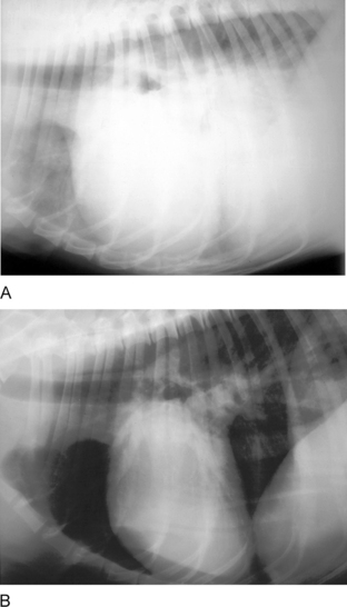
Thoracic radiographs should be examined for generalized cardiomegaly and signs of CHF. Signs of left-sided heart failure include interstitial or alveolar pulmonary edema and moderate to severe left atrial enlargement. Right-sided failure results in pleural effusion, enlarged caudal vena cava, hepatomegaly, and ascites (Figure 38-1).
Echocardiography
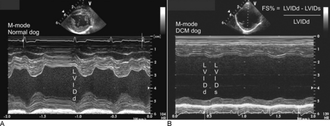
Ventricular and atrial dilation are common with reduced myocardial systolic function (reduced left ventricular fractional shortening percentage [FS%] and ejection fraction). Increased E-point septal separation is also common with ventricular dilation.2 Doppler studies confirm evidence of mitral and tricuspid regurgitation, low-velocity transaortic flow, diastolic ventricular dysfunction, and possible pulmonary hypertension (due to severe left-sided heart failure). Pleural effusion can also be noted along with mild pericardial effusion due to CHF. Remember, most DCM cases with CHF should have moderate to severe atrial enlargement, and low FS% is not pathognomonic for DCM; many normal hearts contract more in apical-to-basilar direction, and this motion is not accounted for by the FS% (Figure 38-2).
Stay updated, free articles. Join our Telegram channel

Full access? Get Clinical Tree


