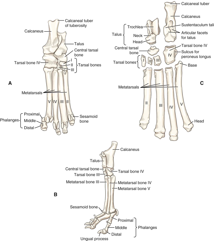
This text is intended for people who already possess knowledge of either veterinary or human anatomy. To assist communication among human rehabilitation and veterinary colleagues, some anatomic terms used for dogs appear in regular print with the analogous terminology for humans in parentheses following the canine term. These comparisons have been minimized, as this is a chapter about canine anatomy and not a chapter about comparative anatomy. Comparative anatomy between dogs and humans has been described in other sources.1–3 The dog stands upright on digits or phalanges of each forepaw or manus and each hindpaw or pes (Figure 5-1). This type of stance is termed a digitigrade stance. The human stands upright on the feet, with the plantar aspect of the feet contacting the floor and adjacent to each other. The upper limbs hang at the sides of the body, palms facing forward. This type of stance is called a plantigrade stance. The main planes of motion for dogs are as follows (see Figure 5-1): • The sagittal plane divides the dog into right and left portions. If this plane were in the midline of the body, this is the median plane or median sagittal plane. • The dorsal plane divides the dog into ventral and dorsal portions. • The transverse plane divides the body into cranial and caudal portions. An axis of rotation for a joint motion is a straight line or rod that is 90 degrees to the plane of motion. For each axis of rotation listed in the next section, the plane of motion around which joint motion occurs can be viewed from Figure 5-1. The axes of rotational joint motion are as follows: • Transverse axis: Sagittal plane motion occurs around an axis of rotation that is directed mediolaterally. • Ventrodorsal axis: Dorsal plane motion occurs around an axis of rotation that is directed ventrodorsally. • Craniocaudal axis: Transverse plane motion, such as rotation of the trunk, occurs around an axis of rotation that is directed craniocaudally. The bones of the dog skeleton and limbs are illustrated in Figures 5-2, 5-3, and 5-4. Bony landmarks on the bones of the limbs are shown in Figures 5-5 through 5-10. The forelimb skeleton consists of the thoracic or pectoral girdle and bones of the forelimb (see Figures 5-5 and 5-6). The size of forelimb bones varies a great deal, because of the greater variation in size for breeds of dogs. The forelimbs bear 60% of the dog’s weight. At the carpus or wrist (see Figure 5-7), there are seven carpal bones. The radial carpal bone is analogous to the fused scaphoid and lunate. There are five metacarpal bones. The first metacarpal is short and nonfunctional. Dogs have many sesamoid bones that are embedded in tendons or near them. Sesamoid bones occur when there are significant changes in directions of pull on tendons in addition to the tensile forces produced during muscle contractions. They allow for constant, biomechanically advantageous alignment of angles of insertion of tendons at their attachment sites, which helps relieve stress on the tendinous insertions for animals that walk on their digits. Dogs are digitigrade animals and bear weight on digits II to V, with the main weight bearing occurring on digits III and IV. The sesamoid bones at the dorsal surface of each metacarpophalangeal joint align the extensor tendons for optimal muscle action. Those on the pad surface of the manus align the flexor tendons. The hindlimb skeleton includes the pelvic girdle, consisting of the fused ilium, ischium, and pubis, and the bones of the hindlimb (see Figures 5-8 and 5-9). The size of hindlimb bones varies a great deal, because of the great variation in size for breeds of dogs. The hindlimbs bear 40% of the dog’s weight. The canine femur is the heaviest4 and largest5 canine bone. In most dogs, it is slightly shorter than the tibia and the ulna and approximately one-fifth longer than the humerus. The average canine angle of inclination or cervicofemoral angle is 144.7 degrees.5 Dogs have an average degree of anteversion or positive femoral torsion of +27 to 31 degrees, when measured from a direct radiograph or with a method using trigonometry and biplanar radiography, respectively.5 The canine femur has a relatively thick and short femoral neck, a caudomedially located lesser trochanter, a prominent lateral greater trochanter, and a relatively short and wide shaft with a narrow isthmus in the middle. The greater trochanter has a craniolateral prominence called the cervical tubercle. Dogs have a third trochanter, which is the attachment site of the superficial gluteal muscle. Canine medial and lateral femoral condyles are equally prominent, but the articular surface of the medial femoral condyle projects more cranially than that of the lateral femoral condyle. The tarsus, or hock, consists of the talus, calcaneus, a central tarsal bone, and tarsal bones I to IV (see Figure 5-10). The talus articulates with the distal tibia and has prominent ridges. At the talocrural joint, two convex ridges of the trochlea of the talus articulate with two reciprocal concave grooves of the cochlea of the tibia. The orientation of the grooves and ridges deviates laterally approximately 25 degrees from the sagittal plane. This deviation allows the hindpaws to pass lateral to the forepaws when dogs gallop.4 The calcaneus is large and serves as the insertion of the common calcaneal tendon. The central tarsal bone lies between the talus and the numbered tarsal bones I to III. Tarsal IV is large and articulates with the calcaneus and metatarsal bones, spanning this entire region. The spine consists of five areas of the vertebral column: the cervical vertebrae and its articulation with the head, thoracic vertebrae, lumbar vertebrae, sacral vertebrae, and the coccygeal vertebrae (Figures 5-11 through 5-14). The number of vertebrae is listed in Box 5-1. All vertebrae, except the sacral vertebrae, remain separate and form individual joints. Four sites with limited motion exist within the canine spine.6 These sites occur at areas where the cranial and caudal articular surfaces are inclined in a nonparallel manner and in different directions. The nonparallel alignment of the articular surfaces markedly restricts joint accessory motions, such as glides. The restricted joint motions and areas resulting from these joint alignments include atlantoaxial motion other than rotation, the cervical (C) 7-thoracic (T) 1 junction, the caudal thoracic region, and the sacrum. The canine atlas, or C1 vertebra (see Figure 5-12), has a transverse foramen in each transverse process, a craniodorsal arch, and right and left lateral vertebral foramina for the passage of cervical spinal nerve 1. The atlas has correspondingly shaped condyles for articulation with the occiput. The canine lateral wings or transverse processes are prominent and easily palpable from the skin surface. The canine axis or C2 has a large spinous process with an expanded arch, a wide body, and large transverse processes (see Figure 5-12). The spinous process is nonbifid. The canine axis is very large relative to the size of other canine cervical vertebrae. The axis has a dens, which projects cranially to allow pivotal motion between the atlas and axis. The condyles are oriented near the transverse plane to allow cervical spine rotation. The C3-C6 vertebrae have nonbifid spinous processes, large and flat spinous processes, caudal and cranial articular surface facets that are narrower than the transverse processes, large transverse processes, and transverse foramina for the passage of vertebral arteries. Caudal and cranial articular surfaces are oriented between the dorsal and transverse planes to facilitate cranial and caudal glides needed for cervical spine flexion and extension. The C7 vertebra has a similar shape, a large prominent nonbifid spinous process, and caudal and cranial articular surfaces, which are oriented nearly craniocaudally. Thoracic vertebrae (see Figure 5-13) have small bodies relative to the size of the entire vertebrae. Canine spinous processes are relatively long. The spinous processes block excessive extension of the thoracic spine. At T10, the size of the body begins to increase and the length of spinous process decreases. The spinous processes are oriented close to the transverse plane. Cranial to T11, the spinous processes project caudally, but caudal to T11, they project cranially. Caudal and cranial articular surfaces are oriented close to the dorsal plane. Lumbar vertebrae (see Figure 5-13) have bodies that are larger than thoracic vertebral bodies. Canine lumbar transverse processes are long and thin, and they project lateroventrocranially. In the cranial lumbar spine, cranial and caudal articular surfaces are oriented between the transverse and sagittal planes, which facilitate lumbar spine flexion and extension. The L7-S1 joint appears to orient between the sagittal and frontal planes to allow more rotation at this intervertebral level. The canine sacrum is relatively narrow and is linked to the pelvis with sacroiliac joints (see Figure 5-14). Caudal (Cd) vertebrae (see Figure 5-14) have distinct bodies and transverse processes. The cranial articular surfaces are similar to those in more cranial vertebrae in shape and location; however, the caudal articular processes are bifid and are more centrally located, whereas articular processes in more cranial vertebrae are located more laterally. Hemal arches are separate bones that articulate with the ventral surfaces of the caudal ends of the bodies of Cd4-Cd6. The hemal arches provide protection for the median coccygeal artery, which is enclosed by the arches. In vertebrae caudal to Cd6 and in relatively the same position as the hemal arches are the paired hemal processes, which extend from Cd7-Cd17 or Cd18. The ribs have vertebral attachments (see Figure 5-11). There are nine pairs of vertebrosternal, or true, ribs and four pairs of vertebrocostal, or false, ribs. The sternum is relatively long and has a manubrium and xiphoid process, with a prominent xiphoid cartilage. The ribs limit overall thoracic spine motion and protect internal organs. The body segments of the forelimb and hindlimb are illustrated in Figures 5-3 and 5-4, respectively, with the major joints and their flexor and extensor surfaces. Body segments are listed and defined in Box 5-1. Types of joints are listed in Box 5-2. Joint motions are named by one body segment approaching or moving away from another body segment or movement of some referenced body landmark. Joint motions are named in the following sections and described (see Figures 5-3 and 5-4) as they refer to the limbs, starting from normal stance. Limb motion is usually described by motion of the joint rather than a body segment. For example, elbow flexion is recommended rather than forearm flexion. Occasionally, body segment motion is used to describe limb motion when motion does not involve axial motion with a joint as a pivot point. For example, rotation of the forelimb might be observable when pronation at the radioulnar joint would be difficult to observe clinically. During flexion, a limb is retracted or folded, a digit is bent, and the back or neck is arched dorsally (i.e., the convex portion of the arch is directed dorsally). In the limbs, flexion motion occurs as the bones on either side of a joint move closer together and the joint angle becomes more acute. Flexion may also be referenced to limb motions involving closing angles during the swing phase of gait. Flexion motions of the limb joints are noted in Figures 5-3 and 5-4. In the spine, flexion occurs as the back or neck arches dorsally (i.e., the convex portion of the arch is directed dorsally). In normal stance, as shown in Figure 5-2, a dog’s spine is flexed at the atlantooccipital and atlantoaxial joints, straight (neither flexed nor extended) in the remainder of the cervical spine, extended at the cervicothoracic junction, slightly lordotic in the thoracic spine, and flexed or normally kyphotic in the lumbar spine. There is either a slightly flexed or extended sacrum on the lumbar spine, depending on the tail posture. The flexed canine lumbar spine is beneficial to running speed. During running, the lumbar spine moves through varying degrees of flexion as running speed changes.
Canine Anatomy
Directional Terms and Anatomic Planes
Directional Terms from Normal Stance (Anatomic Position)

Figure 5-1 Orientation to planes of motion and directional terms for the dog. (From Dyce KM: Textbook of veterinary anatomy, ed 4, St Louis, 2010, Saunders.)
Anatomic Planes
Axes of Rotation
Axes of Rotational Joint Motion
Bones
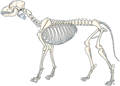
Figure 5-2 Skeleton of a male dog, left lateral view. (From Evans HE: Miller’s anatomy of the dog, ed 4, Philadelphia, 2013, WB Saunders.)
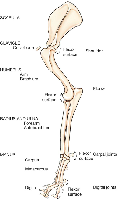
Figure 5-3 Left forelimb skeleton, noting joints and flexor surfaces. (From Evans HE, de Lahunta A: Miller’s guide to the dissection of the dog, ed 7, Philadelphia, 2010, WB Saunders.)
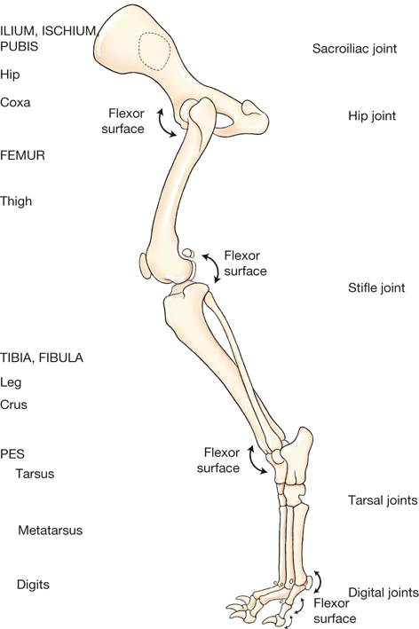
Figure 5-4 Left hindlimb skeleton, noting joints and flexor surfaces. (From Evans HE, de Lahunta A: Miller’s guide to the dissection of the dog, ed 7, Philadelphia, 2010, WB Saunders.)

Figure 5-5 Skeleton of the lateral forelimb of the dog. (Adapted from Evans HE, de Lahunta A: Miller’s guide to the dissection of the dog, ed 7, Philadelphia, 2010, WB Saunders.)
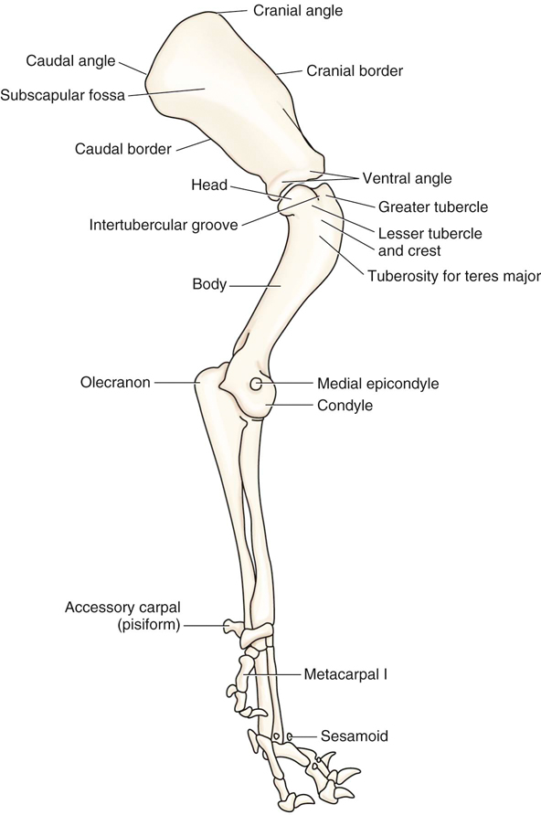
Figure 5-6 Skeleton of the medial forelimb of the dog. (Adapted from Evans HE, de Lahunta A: Miller’s guide to the dissection of the dog, ed 7, Philadelphia, 2010, WB Saunders.)
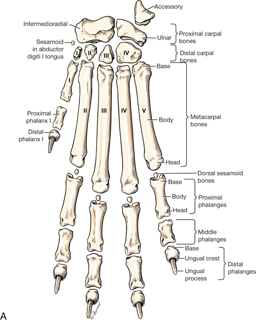
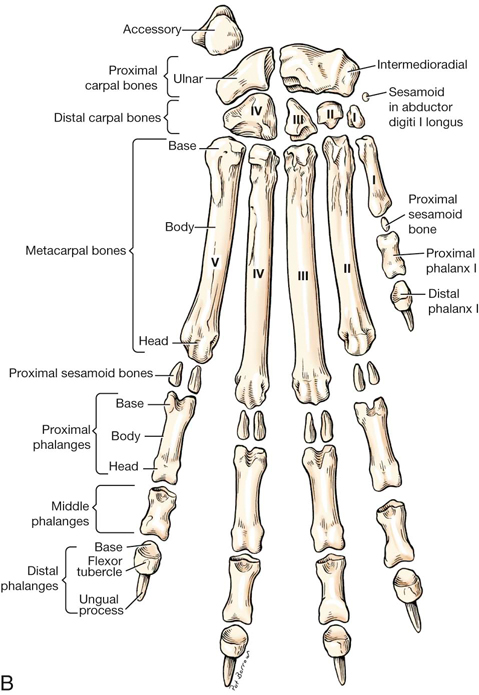
Figure 5-7 Skeleton of the left dorsal (A) and left palmar (B) forepaw of the dog. (From Evans HE, de Lahunta A: Miller’s guide to the dissection of the dog, ed 7, Philadelphia, 2010, WB Saunders.)

Figure 5-8 Skeleton of the lateral hindlimb of the dog. (Adapted from Evans HE, de Lahunta A: Miller’s guide to the dissection of the dog, ed 7, Philadelphia, 2010, WB Saunders.)
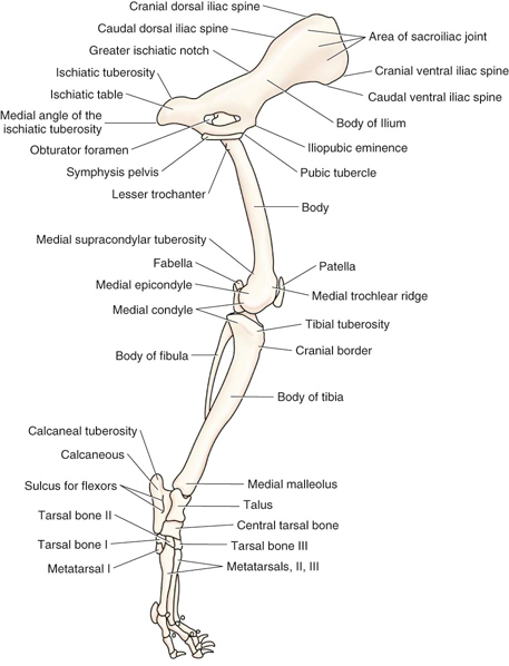
Figure 5-9 Skeleton of the medial hindlimb of the dog. (Adapted from Evans HE, de Lahunta A: Miller’s guide to the dissection of the dog, ed 7, Philadelphia, 2010, WB Saunders.)
Forelimb
Hindlimb
Spine
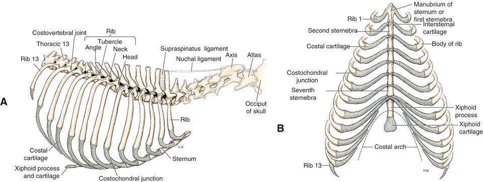
Figure 5-11 A, Identified portions of the axial skeleton cranial to the thirteenth thoracic vertebra. B, Ribs and sternum, ventral view.
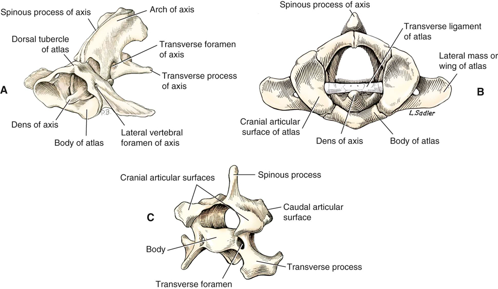
Figure 5-12 Detailed skeletal anatomy of the atlas and axis from a craniolateral view (A), atlas and axis from a cranial view (B), and C5 vertebra from a craniolateral view (C). (A from Evans HE, de Lahunta A: Miller’s guide to the dissection of the dog, ed 7, St Louis, 2010, WB Saunders.)
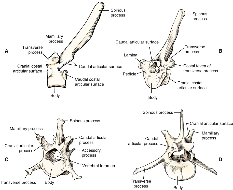
Figure 5-13 Detailed skeletal anatomy of T6 vertebra from a lateral view (A) and craniolateral view (B), L1 vertebra from a craniolateral view (C), and L5 vertebra from a caudolateral view (D).
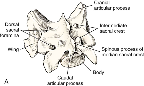
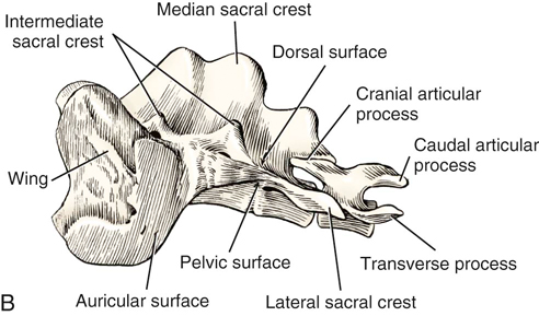
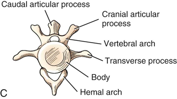
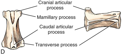
Figure 5-14 Detailed skeletal anatomy of the sacrum from a caudolateral view (A), sacrum and caudal 1 or Cd1 vertebra from a lateral view (B), Cd4 vertebra from a cranial view (C), and Cd6 vertebra from a dorsal view (D). (From Evans HE: Miller’s anatomy of the dog, ed 4, Philadelphia, 2013, WB Saunders.)
Joint Motion
Joint Motion in the Limbs and Spine
Flexion
< div class='tao-gold-member'>
![]()
Stay updated, free articles. Join our Telegram channel

Full access? Get Clinical Tree


Canine Anatomy
Only gold members can continue reading. Log In or Register to continue
