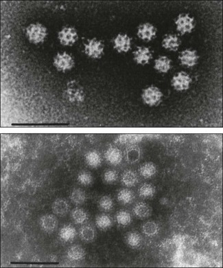
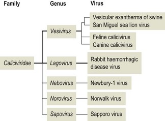
Chapter 53 Caliciviruses derive their name from the Latin word calix meaning cup, which refers to the cup-shaped depressions visible on the surface of the virions in negative-contrast electron micrographs. The virions are icosahedral, non-enveloped and 27–40 nm in diameter with a genome that consists of a single molecule of linear, positive-sense, single-stranded RNA (Fig. 53.1). Replication occurs in the cytoplasm of infected cells with release by cell lysis. Several caliciviruses have not yet been cultivated in vitro. The virions are resistant to ether, chloroform, mild acids and mild detergents. Figure 53.1 (Top) Vesicular exanthema of swine virus showing the cup-shaped depressions that are characteristic of many of the caliciviruses. (Bottom) Enteric calicivirus showing the lack of surface detail that is characteristic of most of these viruses as seen in diagnostic faecal specimens. Negative stain electron microscopy. Bars: 100 nm. Reprinted with permission: Murphy et al., Veterinary Virology Third Edition (1999). Academic Press. Caliciviruses (Table 53.1) are most closely related to picornaviruses and formerly were classified as the genus Calicivirus within the Picornaviridae. The family Caliciviridae is divided into five genera (Fig. 53.2); Vesivirus, Lagovirus, Norovirus, Sapovirus and Nebovirus. Important veterinary viruses in the genus Vesivirus include the prototype virus of the family, vesicular exanthema of swine virus, and feline calicivirus. The Lagovirus genus contains two viruses of lagomorphs, rabbit haemorrhagic disease virus and European brown hare syndrome virus. The human caliciviruses, Norwalk virus and Sapporo virus, are important causes of gastroenteritis. Viruses in the genus Norovirus are also referred to as small, round structured viruses (SRSV). Norwalk virus strains are divided into five genogroups (GI to GV). Bovine noroviruses (GIII) such as Newbury-2 virus as well as porcine noroviruses (GII) and porcine sapoviruses have been described. Other bovine enteric calicivirus isolates have been shown to be distinct and the genus Nebovirus has been created with type species Newbury-1 virus. Hepatitis E virus of man has been removed from the family and placed in a new family Hepeviridae. Table 53.1 Samples rich in virus include vesicular fluid and the overlying flap of epithelium. Diagnostic techniques include ELISA and CFT for antigen detection, immunoelectron microscopy and virus isolation in pig kidney cell lines, with identification by virus neutralization. Primers suitable for both VESV and SMSV have been described for use in RT-PCR (Neill & Seal 1995). A real time assay has also been described (Reid et al. 2007).
Caliciviridae
Virus
Host species
Disease significance
Vesicular exanthema of swine (VES) virus
Pigs
Natural disease not seen since 1956, confined to USA. Acute, contagious, vesicular disease, clinically similar to foot-and-mouth disease. Believed to have resulted from feeding of SMSV-infected sea lion and seal carcasses in swill
San Miguel sea lion virus (SMSV)
Marine mammals, opal eye fish
Causes VES when inoculated into pigs. Cause of cutaneous vesicles and premature parturition in pinnipeds
Feline calicivirus
Domestic and large cats
Upper respiratory tract disease in cats, occurs worldwide. Outbreaks with severe systemic form occasionally described
Rabbit haemorrhagic disease virus
European wild and domestic rabbits
Acute, fatal disease of European rabbits. Physiological resistance in rabbits less than two months of age
European brown hare syndrome virus
European brown hare
Related but distinct from RHDV. Similar disease to RHD, hepatic necrosis and diffuse generalized haemorrhaging. High mortality rate
Canine calicivirus
Dogs
Associated with diarrhoea on occasion
Vesicular exanthema of swine
Caliciviridae
Only gold members can continue reading. Log In or Register to continue

Full access? Get Clinical Tree


