Calcium Channel and β-Blocker Drug Overdose

Chapter 84 Calcium Channel and β-Blocker Drug Overdose


INTRODUCTION


Drugs classified as calcium channel and β-blockers frequently are prescribed for cardiovascular disease management. In humans, these drugs are effective in patients with hypertension, angina pectoris, cardiac arrhythmias, migraines, tremors, and bipolar disorder.1 In veterinary medicine, calcium channel and β-blockers are used to treat cardiac arrhythmias, hypertrophic cardiomyopathy, and hypertension.2-3


Calcium plays a role in many physiologic processes, including impulse formation and conduction, excitation-contraction coupling, and maintenance of vascular tone. Close regulation of intracellular calcium is essential to accomplish these cardiovascular functions.4 There are several types of calcium channels. Calcium channel blockers inhibit only the voltage-sensitive channels, which open in response to voltage changes across the membrane, for example during depolarization. There are three types of voltage-sensitive calcium channels, designated as neuronal (N-type), transient (T-type), and long lasting (L-type). The L-type channels are the most sensitive to the commercially available calcium channel blockers. β-Blockers inhibit the cardiac adrenergic system, modifying the L-type voltage-sensitive channels via a second messenger system.


L-type channels are located in various tissues but are found in highest concentration in the atria, vascular smooth muscle, and skeletal muscle. L-type voltage-sensitive calcium channels are activated as the transmembrane potential of the cell becomes progressively less negative during the upstroke of the action potential (phase 0). They have a prolonged opening time and high conductance, therefore allowing large amounts of calcium to pass rapidly into the cell.5 Calcium channel and β-blockers interrupt calcium flux, leading to decreased intracellular calcium, depressing cardiovascular function.5 Although calcium channel and β-blockers have different mechanisms of action, the physiologic effects, clinical signs, and treatment of toxicity are similar.3


METHOD OF ACTION


Calcium Channel Blockers


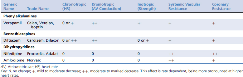
Calcium channel blockers exert most of their effects on cardiac myocytes, pacemaker cells, and vascular smooth muscle. They are classified into three major groups based on their structure, including the phenylalkylamines (e.g., verapamil), the benzothiazepines (e.g., diltiazem), and the dihydropyridines (e.g., amlodipine). Structural differences among the classes are associated with distinct binding sites on the calcium channel, resulting in differing potencies and tissue affinities (Table 84-1). Their structural heterogeneity leads to functional heterogeneity with regard to their vasodilator potency and their cardiac inotropic, chronotropic, and dromotropic effects.1-3


Cardiac Effects


The calcium ion is essential for impulse conductance through the cardiomyocytes. Pacemaker cells of the sinoatrial (SA) and atrioventricular (AV) nodes rely on the inward calcium flux through L-type and T-type channels to initiate a spontaneous diastolic depolarization (phase 4). Calcium channel blockers inhibit inward flow of calcium through the L-type channel, leading to slow SA activity, decreased conduction of impulses through the AV node, and therefore a decrease in heart rate and prolongation of the refractory period.5 The negative chronotropic effect occurs primarily with the phenylalkylamines and benzothiazepines. With some calcium channel blockers, this effect may be attenuated or even abolished because of reflex stimulation of the sympathetic nervous system.2


Calcium plays an important role during excitation-contraction coupling in cardiac and vascular smooth muscles. Within Purkinje cells and myocytes, opening of the L-type calcium channels in response to membrane depolarization increases calcium conductance (phase 2 of the action potential). This inward flow of calcium triggers the release of additional calcium into the cytoplasm from the sarcoplasmic reticulum. Intracellular calcium binds to troponin, changing its conformation and allowing interaction between actin and myosin so that contraction can occur. By decreasing the magnitude and rate of rise of the intracellular calcium concentration, the calcium channel blockers decrease calcium release from the sarcoplasmic reticulum, causing a decrease in the force of contraction.2,5 This negative inotropic effect is seen most commonly with the phenylalkylamines and to a lesser extent with the benzothiazepines.2 Most of the calcium channel blockers have a negative inotropic effect at high doses.3


β-Blockers


There are two types of β-receptors. β1-Receptors are located primarily within the heart and adipose tissue. Stimulation results in increased heart rate, myocardial contractility, AV conduction velocity, and automaticity of subsidiary pacemakers. β2-Receptors are found primarily in bronchial and smooth muscles, where they produce relaxation.4 In human medicine, β-adrenergic blocking agents differ in their ability to block β-receptor types.9 In veterinary medicine the primary drugs are propranolol (β1-receptor and β2-receptor blocker), atenolol (specific β1-receptor blocker), esmolol (specific β1-receptor blocker), and sotalol (β1-receptor and β2-receptor blocker).3


PHARMACOKINETICS


Calcium Channel Blockers


Calcium channel blockers are absorbed rapidly and almost completely from the GI tract but have extensive first-pass metabolism. Times to peak serum concentration are rapid: 20 to 45 minutes for immediate-release forms and 4 to 12 hours for sustained-release formulations and amlodipine besylate (which has a slower absorption rate) in dogs and cats.2 The onset of action varies with the formulation. An animal that bites into and swallows a sustained-release product can show signs within 5 minutes, but one that swallows it whole may not show signs for several hours and may have prolonged toxicity because of slower absorption.10 Tissue distribution is extensive in all classes. Calcium channel blockers are approximately 80% protein bound; therefore interaction with other protein-bound drugs may result in competition for binding sites. Additionally, animals with moderate to severe hypoproteinemia may develop higher blood concentrations.


In humans, calcium channel blockers are metabolized in the liver by oxidative pathways, predominantly by cytochrome P450 CYP3A. Therefore their clearance will be decreased when hepatic function or blood flow is reduced.1 The phenylalkylamines and benzothiazepines can interact with many drugs because they are strong inhibitors of hepatic microsomal enzymes. Similarly, their elimination can be slowed by drugs that inhibit hepatic enzymes (e.g., cimetidine), potentially increasing their cardiovascular effects and producing toxicity.10,11 Elimination half-lives depend on the formulation (i.e., immediate versus sustained release) and in dogs and cats can vary from 2 to 30 hours. Excretion is primarily through urine and, to a lesser extent, bile and feces.2

Stay updated, free articles. Join our Telegram channel

Sep 10, 2016 | Posted by in SMALL ANIMAL | Comments Off on Calcium Channel and β-Blocker Drug Overdose

Full access? Get Clinical Tree

Get Clinical Tree app for offline access