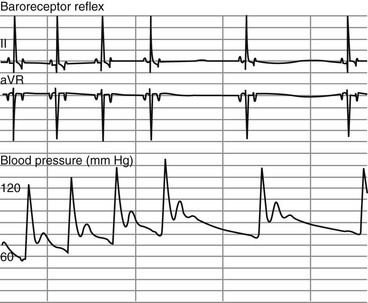
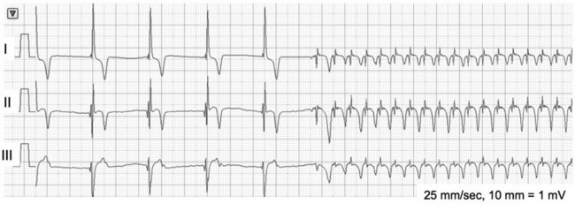
Bradyarrhythmias

Chapter 170


Bradyarrhythmias


Bradyarrhythmias are defined as cardiac rhythms that result in heart rates that are lower than normal for an animal’s signalment and activity. Based on 24-hour ambulatory electrocardiographic (ECG) recordings (Holter monitoring), the average heart rate for healthy dogs is 75 beats/min irrespective of size, whereas the average heart rate for healthy cats is approximately 165 beats/min. However, when rhythms are being evaluated with Holter monitoring in the home, it is equally important to know that a rate of 35 beats/min with sinus pauses of 3 seconds is quite common in a healthy sleeping dog. Importantly, these averages and lower limits may not be applicable to a nervous animal in a stressful environment such as a veterinary hospital. Most clinicians would investigate a heart rate that is persistently less than 140 beats/min in a hospitalized cat and less than 60 beats/min in a hospitalized dog, especially if the rate does not increase with stimulation. When a bradyarrhythmia is diagnosed either in the hospital or via 24-hour Holter monitoring, understanding the cause and significance of the rhythm, and determining optimal management, depends on knowing whether the mechanism is physiologic, iatrogenic, or pathologic.


Physiologic Bradyarrhythmias


Physiologic bradyarrhythmias are those that occur secondary to alteration of extracardiac factors, including body temperature, systemic blood pressure, intrathoracic pressure, intracranial pressure, ocular pressure, gastrointestinal distention, intraabdominal pressure, and pharyngeal or soft palate tension. These bradyarrhythmias are mediated primarily by increases in vagal tone or decreases in sympathetic tone.


A frequently observed physiologic bradyarrhythmia is the development of sinus bradycardia secondary to activation of the baroreceptor reflex. The baroreceptor reflex is an intrinsic physiologic response to an increase in systemic blood pressure that results in an elevation in cardiac vagal tone and a decrease in sympathetic tone (Figure 170-1). The baroreceptors responsible for this response are found primarily in the aortic arch and carotid sinus.


A second physiologic bradyarrhythmia is observed in animals that have brain injury or a disease that increases intracranial pressure and is part of the Cushing’s response. Cushing’s response is a reflex that occurs when the increased hydrostatic pressure of cerebrospinal fluid exceeds mean arterial pressure, which leads to compression of cerebral arterioles that decreases blood flow to the brain. Chemoreceptors in the medulla sense the ischemia-induced increase in carbon dioxide pressure and decrease in pH, resulting in reflex activation of the sympathetic nervous system and peripheral vasoconstriction. This vasoconstriction triggers peripheral baroreceptors to increase vagal tone, and bradycardia results.


Any abnormality in the distribution of the sensory afferent trigeminal nerve fibers can cause an elevation of cardiac vagal tone via activation of the vagal motor nucleus. Space-occupying or infiltrative lesions in the nasal cavity, as well as lesions in the eye or orbital region, may stimulate vagal afferent nerves, which travel via either the ophthalmic, maxillary, or mandibular nerves to the trigeminal ganglion and ultimately activate the vagal motor nucleus. The result of this activation is increased vagal tone and decreased heart rate.


Finally, animals experiencing moderate to severe hypothermia may demonstrate sinus bradycardia secondary to a hypothermia-induced decrease in sinoatrial (SA) node discharge rate.


In each of these examples, a regular or irregular sinus bradycardia is the observed rhythm, but physiologic slowing of atrioventricular (AV) node conduction also can be seen. Although elevated vagal tone is typically the mechanism for physiologic sinus bradycardia, treatment with parasympatholytic agents is not recommended if the animal’s condition and blood pressure are stable; rather, treatment of the underlying problem (e.g., hypothermia, increased intracranial pressure) should be the primary goal. Treatment with atropine or glycopyrrolate may have a dose-dependent pharmacologic effect that is longer than desired. Although the atropine response test has been proposed as a diagnostic test to differentiate physiologic bradycardia from sinus node dysfunction (see the section on sick sinus syndrome later), caution is advised when administering parasympatholytic agents to dogs with potential sinus node dysfunction. The sinus node discharge rate in most dogs with diseased sinus nodes will still increase in response to parasympatholytic agents, which causes some dogs to develop tachycardias that overdrive suppress escape rhythms and can potentially precipitate an asystolic crisis.


Iatrogenic Bradyarrhythmias


Iatrogenic bradyarrhythmias most commonly result from the administration of drugs that decrease SA node discharge rate, AV node conduction, or both. Drugs that slow the heart rate generally are referred to as negative chronotropes. Drug-induced bradycardias most commonly occur in animals given medications for sedation or pain relief and in animals receiving medications used to treat tachyarrhythmias. Centrally acting opioids (e.g., morphine, hydromorphone, butorphanol, fentanyl) and α2-agonists (e.g., dexmedetomidine and medetomidine) can cause a dose-dependent bradycardia. When overdose or adverse response to centrally acting opioids causes a physiologically unstable bradycardia, an opioid antagonist (e.g., naloxone) can be used to reverse the narcotic’s effects. The bradycardic adverse effects of α2-agonist overdose can be reversed with the α2-antagonist atipamezole. Alternatively, anticholinergics such as glycopyrrolate or atropine may be used in either of these situations if symptomatic bradycardia is observed. It should be noted that hypothermia, as commonly occurs in an anesthetic setting, interferes with the effectiveness of anticholinergics in reversing bradycardia.


β-Adrenergic blockers and calcium channel blockers used to treat supraventricular arrhythmias decrease sinus rate and AV node conduction. The latter effect is the rationale for the use of these drugs in controlling the ventricular response rate in atrial fibrillation. Sustained-release diltiazem (2 to 4 mg/kg q12h PO) often is combined with a low dose of digoxin (0.003 mg/kg lean body weight q12h PO) to control ventricular response rate to atrial fibrillation. Digoxin decreases the ventricular rate via its presumed vagomimetic effect on AV node conduction. The authors have had success in controlling ventricular rate without excessive bradycardia using this combination, but the importance of careful monitoring by Holter recordings and serum digoxin concentrations is emphasized (these drugs should be dosed to achieve rate control with serum digoxin concentrations of 0.8 to 1.2 ng/dl and no adverse effects). Importantly, diltiazem’s effect on ventricular rate when given either intravenously or orally is dose dependent, and high doses may induce bradycardia.


Ventricular tachycardia is often treated with sotalol, which has both potassium channel and β-adrenergic blocking effects. The dosage of sotalol required to suppress ventricular arrhythmias (e.g., 2 to 3 mg/kg q12h PO) often decreases the sinus rate by 15% to 20% and may cause a marked increase in the number of pauses exceeding 2 seconds in duration during wakefulness. The duration of pauses during sleep may also be increased by sotalol, and pauses of 5 seconds’ duration are not uncommon in dogs receiving this drug. The bradycardic effects of sotalol most often subside after 4 weeks of treatment, but if sotalol-induced bradycardia is excessive, careful reconsideration of the appropriateness of sotalol therapy and of dosage is recommended.


Finally, general anesthetics have a depressant effect on sinus node function, and in dogs with preexisting conduction system disease, general anesthesia may also suppress ventricular escape activity, leading to asystole. Breeds at risk of sick sinus syndrome (see later) should be monitored carefully during induction and maintenance of anesthesia.


Pathologic Bradyarrhythmias


Pathologic bradyarrhythmias are those nonphysiologic and noniatrogenic bradyarrhythmias that result in a heart rate that is too low to meet the metabolic and perfusion demands of the patient and that therefore result in clinical signs. The clinical signs most commonly observed in patients with pathologic bradyarrhythmias are lethargy, weakness, mental dullness, collapse, and syncope. Pathologic bradyarrhythmias can be categorized loosely into abnormalities of impulse initiation and abnormalities of impulse conduction. Abnormalities of impulse initiation primarily involve an inappropriately low or absent SA node discharge rate and include syndromes such as sick sinus syndrome and persistent atrial standstill. Abnormalities of impulse conduction most commonly consist of a failure of the AV conduction system (AV node, bundle of His, bilateral proximal bundle branches) to conduct a sufficient percentage of depolarizations of sinus origin to the ventricle; the results can include high-grade second-degree and third-degree atrioventricular block (AVB). Third-degree (complete) AVB and sick sinus syndrome are the most common bradyarrhythmias prompting pacemaker implantation in dogs.


Sinoatrial Node Abnormalities


Sick Sinus Syndrome (Sinus Node Dysfunction)


Sinus node dysfunction is a condition in which the SA node intermittently fails to discharge, and this dysfunction may lead to the clinical syndrome of sick sinus syndrome (SSS), in which animals display clinical signs of weakness, lethargy, collapse, or syncope that may or may not be exercise induced. The syncope seen in dogs with SSS is extremely variable with respect to frequency and duration of episodes, and although it is uncommon, dogs with SSS can die if not appropriately treated with pacemaker implantation. Even without a high risk of sudden death, frequent syncope negatively affects quality of life and may prompt unnecessary euthanasia if a client perceives the dog to be suffering.


Although the cause of SSS in dogs is still unclear, mutations in cardiac sodium channels (e.g., SCN5A), connexins, and α smooth muscle heavy-chain proteins; dysfunction of ryanodine receptor 2, which comprises the calcium clock in the SA node; and SA node fibrosis have been identified in people with this disease. SSS is seen most commonly in middle-aged or older small-breed dogs, and canine breeds that are predisposed to SSS include the miniature schnauzer, West Highland white terrier, dachshund, boxer, and cocker spaniel. These breed predispositions suggest a genetic component to the cause of canine SSS. Dogs with SSS may demonstrate sinus bradycardia or a sinus rhythm with periods of sinus arrest. Periods of sinus arrest may be interrupted by junctional or ventricular escape beats or rhythms. Importantly, the escape beats and rhythms in dogs with SSS often are inadequate and delayed, which suggests concurrent dysfunction of secondary (rescue) pacemaker sites in the more distal conduction system. Dogs with SSS also commonly demonstrate a sinus rhythm with intermittent bouts of supraventricular tachycardia that are characterized by positive or negative P waves (bradycardia-tachycardia syndrome) (Figure 170-2). AV conduction disturbances ranging from first- to second-degree AVB of varying severity have been observed, but the prevalence of coexisting AVB is not known. A prolongation of QT interval and sometimes prominent T waves are seen in some dogs with SSS. Abnormal findings may not be apparent on a baseline ECG in dogs with SSS, which highlights the importance of performing 24-hour Holter monitoring or capturing the cardiac rhythm during a clinical event on a loop recorder (event monitor) for definitive diagnosis in many cases. Even if a dog does not have a collapsing episode during the time that Holter monitoring is performed, findings such as a 6- to 7-second sinus pause may strongly suggest a diagnosis of SSS.


< div class='tao-gold-member'>

Stay updated, free articles. Join our Telegram channel

Jul 18, 2016 | Posted by in PHARMACOLOGY, TOXICOLOGY & THERAPEUTICS | Comments Off on Bradyarrhythmias

Full access? Get Clinical Tree

Get Clinical Tree app for offline access