Chapter 45 Bradyarrhythmias and Conduction Abnormalities
INTRODUCTION
Conduction abnormalities may or may not produce an arrhythmia. The term arrhythmia literally means “no rhythm”. Because of this, some prefer the term dysrhythmia. However, arrhythmia is the more commonly used term and is used here to describe any abnormal heart rhythm. Mechanisms of arrhythmias primarily include disorders of cardiac electrical impulse conduction and electrical impulse formation. Conduction abnormalities commonly result in conduction delays and blocks but can also contribute to formation of ectopic tachyarrhythmias (premature depolarizations) by producing a substrate for reentry. Abnormalities of impulse formation produce both bradyarrhythmias and tachyarrhythmias.1
DISORDERS OF ELECTRICAL IMPULSE CONDUCTION
Almost all cardiac tissues depolarize during systole and help conduct the depolarization wave from site to site. Disease of cardiac tissue can result in regions of conduction delay or conduction block. Conduction delays or blocks can result in bradyarrhythmias or tachyarrhythmias.1
CONDUCTION ABNORMALITIES LEADING TO BRADYARRHYTHMIAS
Conduction abnormalities that lead to bradyarrhythmias are due to conduction delays or conduction blocks within the specialized conduction system. Conduction starts in the tissues surrounding the sinus node and terminates in the Purkinje network in the ventricles. Slowed conduction from the sinus node to the internodal tracts (first-degree sinoatrial block) does not cause any perceptible abnormality on an electrocardiogram (ECG) because it occurs before the P wave is inscribed. An intermittent conduction block in this region (second-degree sinoatrial block) results in the heart rhythm stopping, usually only for one beat, because of the lack of a P-QRS-T complex on the ECG. Complete blockage of conduction from the sinus node to the internodal tracts and atria theoretically results in atrial standstill and forces the atrioventricular (AV) node to take over the pacing function of the heart at a slower rate. In reality, other regions of automaticity (e.g., tissue around the coronary sinus) in the atria probably take over the function of the sinus node in this situation.1
Slowed conduction through the AV conduction system results in a prolongation in the PR interval (first-degree AV block). This conduction delay can occur in the proximal AV bundle, the AV node, the bundle of His, or the bundle branches (if both the left and right bundle branches are affected). It theoretically may also occur in the internodal tracts. An intermittent complete block of conduction results in the intermittent loss of a QRS-T complex (second-degree AV block). Third-degree AV block occurs when conduction is completely blocked through the AV node, bundle of His, or both bundle branches. Complete block of conduction through the internodal tracts is also reported to produce complete AV block (third-degree AV block) in dogs.2
DISORDERS OF IMPULSE FORMATION
Disorders of impulse formation encompass enhanced or depressed impulse formation by abnormal pacemaker cells and abnormal impulse formation by cells that are not normally automatic.1
DEPRESSED NORMAL AUTOMATICITY
A depression in normal automaticity results in a decrease in the discharge rate of an automatic site. This can be due to disease of the automatic tissue or depression of automatic tissue as a result of diverse influences, such as the parasympathetic nervous system, electrolyte disturbances (e.g., hyperkalemia), endocrine abnormalities (e.g., hypothyroidism), and hypothermia. To be manifested as a bradyarrhythmia, the sinus node must be affected, either by itself or in combination with subsidiary pacemaker sites. For example, if the automatic cells within the AV node are suppressed by stimulation of the left vagus nerve such that their inherent rate decreases from 50 to 30 beats/min, but the sinus node continues to depolarize at a rate of 100 beats/min, the AV nodal cell depression will never be identified because the faster sinus nodal rate continues to control the heart rate. Normal automaticity can also be depressed by disease. Sick sinus syndrome is a disease in which the sinus node tissue is diseased and destroyed. When most of the sinus node tissue is destroyed, it loses its ability to produce depolarizations automatically.1
SPECIFIC BRADYARRHYTHMIAS
Sinus Bradycardia
Sinus bradycardia is a regular rhythm that originates in the sinus node but at a rate that is too slow for a given situation. A sinus rate less than 60 beats/min in an awake dog in an examination room is generally considered too slow. However, the sinus rate can be as slow as 20 beats/min in a normal dog that is sleeping. A heart rate less than 100 to 120 beats/min in a cat is generally too slow, although it may produce no clinical signs. Sinus bradycardia is an uncommon rhythm disturbance in clinical veterinary practice. It is identified most commonly during an anesthetic overdose. Other causes include increased vagal tone (athletic training, increased intracranial pressure, severe gastrointestinal (GI) or respiratory disease), sick sinus syndrome, hypothermia, severe hypothyroidism, and administration of parasympathomimetic or sympatholytic drugs, such as xylazine, digoxin, and β-blockers. Sick sinus syndrome is discussed later in this chapter. 1,3
Treatment
Treatment of sinus bradycardia is unnecessary unless clinical signs are evident, and it depends on the underlying cause. If drug administration is precipitating the problem, it should be discontinued. If the problem is peracute, such as in a patient under anesthesia, atropine should be administered in an attempt to increase the heart rate. If this is unsuccessful, a β-adrenergic agonist, such as isoproterenol, can be administered; however, close monitoring for hypotension is recommended. Conscious patients that have no apparent underlying cause should be given atropine (0.04 mg/kg SC or IV) and an ECG repeated 30 minutes or 10 minutes later, respectively. In a dog with a normal sinus node, the heart rate should increase to 140 to 200 beats/min. If a normal response is identified, the diagnosis of increased vagal tone is made. If the dog is symptomatic, chronic anticholinergic therapy can be initiated. If there is no response or if the response is only partial (heart rate increases to 70 to 130 beats/min), sick sinus syndrome is most likely present.1
Sinoatrial Block
Sinoatrial block occurs when the tissue surrounding the sinus node fails to conduct the depolarization to the atria and ventricles. Some, but not all, depolarizations are conducted in a second-degree sinoatrial block. This is the most commonly diagnosed type of sinoatrial block. Second-degree sinoatrial block is diagnosed on an ECG when a pause occurs after a sinus beat and the interval between beats is an exact multiple (e.g., 2 or 3 times) of the normal P-P interval. This indicates that the sinus node is most likely depolarizing at its normal rate, but the depolarization is being blocked intermittently from conducting to the atria and internodal tracts. Consequently, no P waves and no QRS-T complexes are produced.1
Sinus Arrest
Sinus arrest is a cessation of sinus node activity for a short period. Although sinus arrest commonly is described as a pause in the sinus rhythm that lasts for more than two normal R-R intervals, this can also be seen with severe sinus arrhythmia. Consequently, there is a “gray zone” between severe sinus arrhythmia and sinus arrest in the dog.1 Sinus arrest in dogs most commonly is due to either sinus node dysfunction or increased vagal tone. Sinus node dysfunction usually is due to end-stage sinus node disease and is commonly called sick sinus syndrome. Sinus node dysfunction in dogs can produce other arrhythmias, including sinus bradycardia and the so-called tachycardia-bradycardia syndrome. A supraventricular tachycardia can sometimes be seen with sick sinus syndrome, presumably because of reentry in or around the diseased sinus node. However, sinus arrest is the most common manifestation of end-stage sinus node disease in dogs.4 Increased vagal tone can also produce periods of sinus arrest and can, on occasion, produce pauses long enough to cause syncope. Vagal tone can be increased secondary to chronic respiratory disease or secondary to systemic disease (e.g., GI disease).1
Escape Beats
Sinus arrest can last for a short period (<1 second in a dog) and be terminated by the sinus node depolarizing again. Sinus arrest can also last long enough that a subsidiary pacemaker, such as the AV node, takes over the heart rhythm. When the AV junction or the Purkinje fibers take over the heart rhythm, QRS complexes occur after a pause or occur at a rate between 20 and 60 beats/min. Depolarizations that occur after a pause are called escape beats, and the slow rhythms are called escape rhythms. Escape beats normally originate either from the AV junctional region or from Purkinje fibers. Because the rates for these two sites differ, one can determine the origin of an escape beat by determining the rate at which it fires.
Sick Sinus Syndrome
Dogs with diffuse conduction system disease or with increased vagal tone to both the sinus node and the AV node may have more prolonged periods of sinus arrest because the subsidiary pacemakers are either suppressed or dysfunctional. If a period of sinus arrest lasts for more than approximately 6 seconds, weakness and syncope will occur.4
Treatment
Dogs with sick sinus syndrome generally require the implantation of an artificial pacemaker to prevent syncope. Occasionally a dog that is partially responsive to atropine administration can be treated chronically with an anticholinergic or a sympathomimetic agent. However, the disease usually progresses to the point that the arrhythmia becomes unresponsive to drug therapy and pacemaker implantation is required.
Dogs with vagally induced sinus arrest require therapy if clinical signs of episodic weakness or syncope occur. Anticholinergic or sympathomimetic therapy should be tried initially. Anticholinergic agents that can be administered on a long-term oral basis include atropine, isopropamide, prochlorperazine plus isopropamide (Darbazine), and propantheline. Atropine tablets are no longer manufactured. Isopropamide and propantheline are weak anticholinergic agents compared with atropine and generally are not as effective. Anticholinergic agents can produce side effects, including mydriasis and constipation.
Alternatively, a sympathomimetic agent can be administered. Terbutaline and albuterol syrup are the two choices. Sympathomimetic drugs act indirectly by counteracting the effects of increased vagal tone. They can produce side effects, including hyperactivity and tachycardia. Dosage adjustment may reduce the side effects while maintaining efficacy. Dogs that have intolerable side effects or that are unresponsive to medical therapy should have a pacemaker implanted. The pacemaker will prevent clinical signs that occur secondary to sinus arrest.1
ATRIAL STANDSTILL
Definition, Causes, and Electrocardiographic Findings
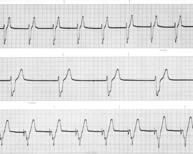
Atrial standstill is the rhythm diagnosis when no P waves are visible on the ECG and atrial fibrillation is not evident (Figure 45-1). Atrial standstill occurs when the atrial myocardium is unable to depolarize. This occurs for two broad general reasons: (1) either the atrial muscle is destroyed by disease or (2) the serum potassium concentration is increased to a level at which the resting membrane potential of atrial cells is so low (i.e., closer to zero) that they no longer depolarize.

Figure 45-1 Lead II electrocardiogram tracings from a dog that was presented for being lethargic for 1 month and that had had ascites for the previous 2 weeks. The heart rate was 50 to 60 beats/min on auscultation. There are no P waves visible in the top and middle tracings. The top tracing is recorded at 25 mm/sec, and the middle trace is recorded at 50 mm/sec paper speed. The ventricular rate is 55 beats/min and regular. These features are characteristic of atrial standstill. The QRS complexes are wide and bizarre in appearance. The configuration of the QRS complexes indicates that the escape focus in this dog is either in Purkinje fibers (ventricular escape beats) or in the atrioventricular junctional tissue and not conducted in the right bundle branch (nodal escape rhythm with a right bundle branch block). Because the rate is consistent with a nodal escape rhythm, the escape focus is most likely in the atrioventricular junctional tissue. The bottom tracing was recorded at 50 mm/sec after pacemaker implantation. The generator was set at a rate of 100 beats/min. A sharp deflection precedes each QRS complex. This is a so-called pacemaker spike that occurs when the generator produces its electrical signal. The pacemaker spikes are exactly 0.6 second apart, indicating that the set rate (100 beats/min) is being produced. The QRS complexes are wide and bizarre in appearance, because the wave of depolarization originates within myocardium and must conduct from muscle cell to muscle cell. (1 cm = 1 mV.) (From Kittleson MD: Diagnosis and treatment of arrhythmias [dysrhythmias]. In Kittleson MD, Kienle RD, editors: Small animal cardiovascular medicine, St Louis, 1998, Mosby.)
From Kittleson MD: Diagnosis and treatment of arrhythmias [dysrhythmias]. In Kittleson MD, Kienle RD, editors: Small animal cardiovascular medicine, St Louis, 1998, Mosby.

Stay updated, free articles. Join our Telegram channel

Full access? Get Clinical Tree

