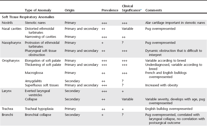
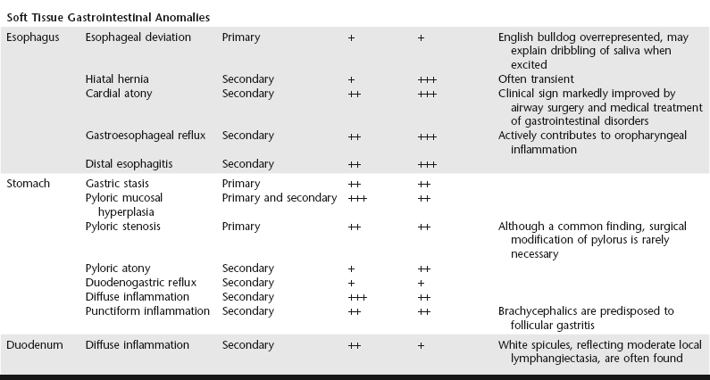
Brachycephalic Airway Obstruction Syndrome

Chapter 156


Brachycephalic Airway Obstruction Syndrome


Decades of genetic modification of brachycephalic breeds in the pursuit of ever flatter facial features have led to the progressive development of achondroplasia. This congenital and hereditary disorder leads to arrested elongation of nasal bones and results in relative hyperplasia of the soft tissues, with numerous deleterious consequences for the respiratory, gastrointestinal, and cutaneous systems. Since a close interaction has been established between respiratory and gastrointestinal disorders, the latter are considered an integral part of brachycephalic airway obstruction syndrome (BAOS).


In addition to stenotic nares and elongated soft palate, numerous other anomalies have been identified in these dogs. They may be congenital (primary) or acquired (secondary) (Table 156-1). The presence and severity of these anomalies vary from one dog to another and among the affected canine breeds. Although the clinical incidence of each entity may be expressed individually, each abnormality participates to a greater or lesser extent in an “engorgement” of the upper airways within a nondistensible cranial space.


A common pathophysiologic pathway seemingly can be identified for the respiratory and gastrointestinal anomalies of dogs with BAOS: gastroesophageal disorders and defective emptying of the upper gastrointestinal tract may aggravate the respiratory signs by encumbering the pharyngeal region and stimulating inflammation. Conversely, persistently increased respiratory effort promotes gastroesophageal reflux and other gastrointestinal conditions commonly described in these dogs. There is a clinical correlation between the severity of the respiratory disorders and the severity of gastrointestinal disorders that can be identified in the patient history.


Clinical Signs


The clinical presentation is dominated by respiratory signs, which are always present with inspiratory or mixed inspiratory-expiratory effort and noise. Intolerance of exercise, stress, and heat is characteristic. Snoring with stertor or stridor, coughing, and throat clearing are also reported. Episodes of cyanosis and syncope are commonly described in severely affected dogs. The progression of the clinical signs is notable, with gradual worsening over the months and with passing summers. Owners often consult a veterinarian during the dog’s first summer after puppyhood or during an acute period of respiratory distress, although some owners consider these signs “normal” for the breed.


The prevalence of gastrointestinal disorders is significant in dogs with upper airway obstruction and can be the principal reason for consultation. A study of 73 cases (Poncet et al, 2005) showed a high incidence of concurrent gastrointestinal disorders in brachycephalic dogs brought for treatment primarily of respiratory disorders. The clinical signs most commonly described by the owners include vomiting (of gastric juices or food, indicative of gastric retention), regurgitation (sometimes linked to exertion, but very commonly occurring when the dog becomes excited), eructation, ptyalism and repeated swallowing, and ingestion of grass or other form of pica.


Diagnostic Approach


Various complementary examinations are required in the global management of obstructive airway syndrome. The purpose of these examinations is to determine an overall profile of the anomalies present, which is essential for planning treatment and determining prognosis. Cervical and thoracic radiographs are obtained to detect bronchopneumonia from food inhalation, bronchiectasis, pulmonary edema, tracheal hypoplasia, or deformation of the cardiac silhouette. Echocardiography is useful if heart disease is suspected. Computed tomography or magnetic resonance imaging can be helpful for modelling the soft palate and exploring the nasal cavities.


Endoscopy of the upper respiratory and gastrointestinal tracts provides a good overview of the lesions. Retrograde endoscopy of the nasopharynx enables documentation of choanal obstruction, notably via expansion of the ethmoidal turbinates. Antegrade rhinoscopy can be used to identify intranasal obstruction. Esophageal lesions commonly observed in brachycephalic dogs include esophageal deviation and distal esophageal erosions and inflammation secondary to chronic reflux. Other gastrointestinal findings include gastric fundal and antral inflammation (and notably follicular gastritis), gastric retention (evidenced by persistence of partially digested food despite prolonged consumption of a fluid-only diet), reduced pyloric diameter with hypertrophic mucosal folds, and gastroduodenal inflammation.


Endoscopy of the upper gastrointestinal tract enables visualization of gastrointestinal lesions and collection of gastric and duodenal biopsy samples; these are essential in dogs with gastrointestinal signs, because there is a poor correlation between the macroscopic and histologic appearance of the lesions. A prospective study of gastrointestinal lesions in 73 brachycephalic dogs (Poncet et al, 2005) revealed marked histologic lesions in 98% of cases in which biopsy samples were available. In this study a significant correlation was also established between the severity of the respiratory obstruction and the severity of gastrointestinal disorders.

< div class='tao-gold-member'>

Only gold members can continue reading. Log In or Register to continue

Stay updated, free articles. Join our Telegram channel

Jul 18, 2016 | Posted by in PHARMACOLOGY, TOXICOLOGY & THERAPEUTICS | Comments Off on Brachycephalic Airway Obstruction Syndrome

Full access? Get Clinical Tree

Get Clinical Tree app for offline access