Blood Film Evaluation

Chapter 122 Blood Film Evaluation


KEY POINTS


BLOOD FILM EVALUATION


A systematic approach to blood film evaluation is critical to obtaining accurate and complete results. Blood films should first be scanned at low magnification (10× to 20×). All three areas of the blood film (the body of the film near the drop, the monolayer where red blood cells are seen as individuals and may touch but do not overlap, and the feather edge which is most distant from the drop) should be examined. Scan for clumping of cells (platelets, white blood cells, or red blood cells), rouleaux (orderly stacking of red blood cells), and agglutination (three-dimensional clumping of red blood cells). Any atypical cells or organisms (parasites) should be noted.3


Cell morphology is assessed in the monolayer. Even at scanning magnification, one can estimate total white blood cell count and differential. Typical appearance of each leukocyte cell line (neutrophil, eosinophil, monocyte, and lymphocyte) can be assessed. Individual red blood cells can be evaluated for evidence of anisocytosis, polychromasia, and poikilocytosis.


After the slide has been scanned, the entire process should be repeated at oil immersion magnification (100×). Again, most of the evaluation is done in the monolayer. Leukocytes are evaluated for morphologic detail. The presence of neutrophil toxicity, reactive lymphocytes, and inclusions in neutrophils and monocytes are noted. A left shift, which can be recognized at scanning magnifications, is confirmed at oil immersion magnification.


Red blood cell changes suspected at scanning magnification (anisocytosis, polychromasia) also can be confirmed. Poikilocytosis is best assessed at oil immersion magnification. The various types of poikilocytes (spherocytes, schizocytes, acanthocytes, dacryocytes) can all be recognized. Red blood cell inclusions, such as Heinz bodies, and red blood cell parasites, such as Mycoplasma and Babesia, can be observed.


Oil immersion magnification is best for evaluating platelet numbers. In general, in the monolayer there should be 8 to 15 platelets per 100× field. This number can be reduced significantly, despite a normal platelet count, if platelet clumps are present.


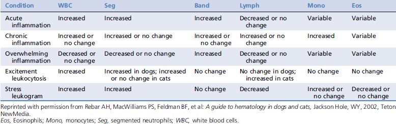
WHITE BLOOD CELL RESPONSES1,3


White blood cell responses are among the best laboratory the indicators of the general health status of critically ill patients. Many emergent and critically ill patients suffer from inflammatory diseases. Inflammation is usually indicated by the leukogram. Signs of inflammation include a left shift, monocytosis, or persistent eosinophilia. General patterns of leukocyte responses are found in Table 122-1.


Left shifts (increased numbers of immature neutrophils in the blood) can be further classified as regenerative or degenerative, depending upon whether total neutrophil counts are increased or decreased. Increased numbers of mature neutrophils in conjunction with a left shift is a regenerative left shift (Color Plate 122-1). Regenerative left shifts indicate that bone marrow production of neutrophils is keeping pace with tissue demand, which results in a favorable prognosis for the patient. In contrast, degenerative left shifts have normal or decreased numbers of mature neutrophils, indicating that marrow production is not keeping pace with tissue demand. This in turn leads to a guarded prognosis for the patient. A special form of degenerative left shift occurs when total neutrophil counts are increased, but over 50% of the neutrophils are immature.


Monocytosis (Color Plates 122-1 and 122-2) not only indicates inflammation, but is also an indicator of demand for phagocytosis or tissue necrosis. Monocytosis can occur acutely or chronically depending on the inciting cause of the inflammation. Acute systemic diseases such as histoplasmosis and toxoplasmosis have been shown to cause monocytosis in as little as 8 to 12 hours after infection. A mild increase in circulating monocytes may also be secondary to a stress leukogram.


Persistent eosinophilia usually indicates a systemic hypersensitivity reaction. Causes include systemic parasitic infections (heartworm infections, migrating parasitic larvae), systemic mastocytosis, allergic reactions, feline asthma, and allergic gastroenteritis. In addition to mastocytosis, some other neoplastic diseases, most notably lymphoproliferative disorders, may be associated with systemic hypersensitivity reactions and persistent eosinophilia.


A persistent eosinophilia is also a feature of hypoadrenocorticism (Addison’s disease). In this circumstance there is generally a lymphocytosis and a lack of a mature neutrophilia. Patients with Addison’s disease may arrive in a state of collapse or severe weakness and depression. In these instances, a complete blood count is always warranted.


Other lymphocyte responses can also be quite informative in patients with inflammatory disease. Mild to moderate lymphopenia (counts from 750 to 1500 cells/μl) is usually the result of high circulating corticosteroids (stress response). Marked lymphopenia may be caused by stress alone, but other causes of lymphopenia should also be considered. These include any disease state that interferes with the normal circulatory pattern of lymphocytes (lymph to blood to tissues to lymph). Possibilities include lymphoma, chylous effusions, and lymphedema. Chylous effusions and lymphedema are generally characterized by low plasma and serum protein levels.


White blood cell morphology can be as informative in patients with inflammatory conditions as white blood cell numbers.1-3 Neutrophils should be assessed for toxicity. The most common feature of toxicity in neutrophils is foamy basophilia of the cytoplasm (Color Plate 122-3). Dohle bodies, or intracytoplasmic basophilic precipitates of ribonucleic acid (RNA) (Color Plate 122-4), also represent toxic change. The presence of Dohle bodies indicates significant toxicity in dogs but is far less important in cats, in which they can be seen even in healthy animals. Other features of toxicity include the presence of giant neutrophils and aberrant nuclear shapes such as ring forms.


Neutrophil toxicity indicates that circulating toxins are interfering with neutrophil development in the bone marrow or that neutrophil production has been accelerated in the bone marrow as a result of increased peripheral demand. Toxicity is most commonly associated with bacterial infections but can be observed in other inflammatory diseases as well. For example, toxicity can be seen following extensive tissue necrosis (as sometimes is seen with severe trauma).


Following the appearance and disappearance of neutrophil toxicity can be important for the treatment of the critically ill patients. The appearance of toxicity may indicate that the patient’s condition is worsening. Resolution of toxicity is often an indicator of improvement. However, the assessment of toxicity may be prone to subjective evaluation unless the same person reviews the slide daily, making interpretation of changes over time more challenging.


The presence of reactive lymphocytes should also be noted. Reactive lymphocytes are generally larger than normal, with increased amounts of basophilic cytoplasm and large nuclei containing finely granular chromatin. Reactive lymphocytes are antigen stimulated and indicate that the immune system has been engaged.1-3


Monocytes and neutrophils should be examined closely for inclusions. A variety of infectious disease agents, including Ehrlichia, Histoplasma, Hepatozoon, and Leishmania, can sometimes be observed in circulating phagocytes. Erythrophagocytosis is sometimes seen with certain red blood cell disorders such as immune-mediated hemolytic anemia (IMHA).

Stay updated, free articles. Join our Telegram channel

Sep 10, 2016 | Posted by in SMALL ANIMAL | Comments Off on Blood Film Evaluation

Full access? Get Clinical Tree

Get Clinical Tree app for offline access