Bleeding Disorders

Chapter 118 Bleeding Disorders


PHYSIOLOGY OF HEMOSTASIS


The hemostatic system is complex; the numerous components function as an interrelated continuum to prevent excessive bleeding or thrombosis following injury. For the emergency clinician, however, it is convenient and sufficiently accurate to divide hemostasis into three component parts: primary hemostasis, secondary hemostasis, and fibrinolysis.


Primary hemostasis involves interactions between the platelets and the endothelium, culminating in the formation of the primary hemostatic plug, which constitutes a temporary seal over the injured vessel. At the site of vascular injury, platelets adhere to subendothelial collagen, mediated largely by von Willebrand factor (vWF) and membrane glycoproteins.1,2 Following adherence, the platelets undergo conformational changes and activation. This includes the release of bioactive agonists that stimulate platelet aggregation and recruitment.1,2 Aggregated platelets constitute the primary hemostatic plug and provide a stimulus and framework for secondary hemostasis. Defects in primary hemostasis may be due to disorders of the platelets or the vasculature. Platelet disorders can be quantitative (thrombocytopenia) or qualitative (thrombopathia). Vasculopathies result in excessive fragility or abnormal endothelium-platelet interactions.


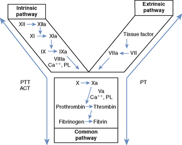
Secondary hemostasis comprises a series of enzymatic reactions, culminating in the cleavage of plasma fibrinogen to form fibrin, which stabilizes the primary hemostatic plug.1,3 All coagulation factors participating in secondary hemostasis are produced in the liver. Factors II (thrombin), VII, IX, and X are proenzymes that undergo a vitamin K–dependent modification before secretion from hepatocytes.1,3 Classically, two pathways of coagulation activation are recognized: an intrinsic and an extrinsic pathway (Figure 118-1). The intrinsic pathway is surface activated and operates strictly with the components present in the blood, whereas the extrinsic pathway requires a tissue factor for activation. These two pathways converge in a final common pathway of thrombin generation and fibrin formation. Defects of secondary hemostasis may be quantitative or qualitative coagulation factor disorders, or both.


The fibrinolytic system consists of plasminogen and all substances that convert it to its active form, plasmin. Plasmin is responsible for dissolution of the fibrin clot. Dissolution results in the production of various fragments called fibrin split products (FSPs), also known as fibrin degradation products. FSPs ultimately are removed from the circulation by the liver (half-life approximately 9 to 12 hours).4 FSPs interfere with platelet function and inhibit thrombin, thus contributing to a bleeding tendency.1,4 The breakdown of fibrinogen can also increase measured levels of FSPs or FDPs.


EMERGENCY APPROACH TO THE BLEEDING PATIENT


The patient may present for bleeding that is evident to the owner. It may also present for signs related to anemia from ongoing hemorrhage, or signs due to acute bleeding that compromises organ function or hemodynamics. Patients in an anemic crisis are depressed or moribund, with marked pallor, tachypnea, tachycardia, and bounding pulses. If bleeding has been gradual and there has been sufficient time for compensatory fluid shifts, the patient may be weak but hemodynamically stable. If anemia is due to substantial acute blood loss, signs of hypoperfusion predominate. Hemorrhage into the brain, spinal cord, myocardium, or lungs can result in acute organ compromise without significant anemia or shock.


A primary survey should be performed in any emergency patient. This is the initial rapid assessment of vital organ systems to determine if a life-threatening situation exists. Hypovolemic shock, an anemic crisis, and pulmonary or brain hemorrhage constitute life-threatening situations in the bleeding patient. Venous access should be established without delay and blood collected from the catheter for a minimum database, including a packed cell volume (PCV) and total protein (TP). In the bleeding animal, both PCV and TP are usually decreased. In animals with acute hemorrhage, however, the PCV may be normal or elevated as a result of inadequate time for fluid redistribution or compensatory splenic contraction. A low TP in a hypovolemic patient is suggestive of acute blood loss, regardless of the PCV. Additional blood samples should be collected before initiating therapy to avoid treatment-induced changes in laboratory parameters. These should include a blood smear, serum, ethylenediaminetetraacetic acid (EDTA) plasma sample, and citrated plasma sample for later testing.


Following sample collection, therapy should be initiated to stabilize the patient. Initial stabilization of the bleeding patient involves (1) control of hemorrhage, when possible; (2) blood transfusion, if anemia is significant; and/or (3) blood volume replacement, when hypoperfusion is present (see Chapters 65 and 66, Shock Fluids and Fluid Challenge and Transfusion Medicine, respectively). In animals with hemorrhagic shock, the most life-threatening problem is hypoperfusion. Initial therapy therefore should involve aggressive fluid therapy (isotonic crystalloids with or without synthetic colloid fluids) until blood is available. There is no justification for withholding fluid therapy in the anemic patient. Hypoperfusion will only exacerbate the tissue hypoxia.


The patient with a suspected hemorrhagic tendency should be kept quiet. Subcutaneous injections should be avoided, when possible, and venipuncture performed only when required for platelet enumeration. Venipuncture sites should be held with manual pressure for atleast 5 minutes. An intravenous catheter usually can be placed safely and is used to collect all other blood samples. The patient should be monitored closely for evidence of ongoing or recurrent hemorrhage, including evaluation of perfusion, respiratory, and neurologic status, mucous membrane color, and PCV and TP, as well as monitoring of blood pressure and electrocardiogram. Following initial stabilization, every effort should be directed toward establishing a rapid diagnosis.


DIAGNOSTIC APPROACH


Three initial questions must be answered: (1) Is the bleeding a result of local factors or does the patient have a generalized bleeding disorder? (2) If a systemic bleeding disorder does exist, what is the nature of the defect (primary or secondary hemostasis)? (3) Is the defect congenital or acquired? These questions can usually be answered based on the history, physical examination, and screening laboratory tests.


History


The importance of a thorough and detailed history cannot be overemphasized. Information should be sought regarding past or present bleeding episodes that prompted presentation or a history of recent trauma. In some cases, bleeding may not be apparent to the owner. Lameness may result from hemarthrosis and dyspnea from intrapulmonary hemorrhage. The owner should be questioned regarding any evidence of bleeding in other sites that would indicate a systemic bleeding disorder.


The signalment of the patient may be informative. Severe inherited disorders are generally apparent within the first 6 months of life. Milder forms, such as von Willebrand disease (vWD), may not be diagnosed until surgery, trauma, or concurrent disease precipitates bleeding. Acquired hemostatic anomalies are seen more commonly in mature animals. It can be difficult to differentiate between a mild inherited defect and a newly acquired disorder. A history of repeated bleeding episodes suggests a possible inherited coagulopathy. Acquired disorders can occur in any breed. Some breeds, however, appear to be more prone to certain disorders (e.g., immune-mediated thrombocytopenia [IMTP] in Cocker Spaniels). Inherited disorders show a much higher breed predilection.


The clinician should try to determine whether bleeding episodes occurred spontaneously or were precipitated by injury or surgery. Some inherited disorders (e.g., hemophilia) and many acquired disorders (e.g., thrombocytopenia, vitamin K deficiency) produce spontaneous bleeding, whereas milder forms of these diseases and other conditions (e.g., vWD, factor VII deficiency) more commonly require some form of trauma to make the clotting impairment clinically apparent. The assessment of response to trauma may also enable the clinician to date the onset of the disorder. A patient that has tolerated surgery is unlikely to have a severe inherited bleeding disorder.


The history should include detailed enquiries about previous illnesses and past and present medications. Many systemic diseases can compromise hemostasis and precipitate clinical bleeding, particularly in a patient with an already compromised hemostatic mechanism. Numerous drugs have been associated with thrombocytopenia, thrombopathias, and coagulopathies. Live-virus vaccines and certain drugs can cause thrombocytopenia 3 to 10 days post administration. A travel history may elucidate exposure to infectious diseases. Specific enquiries about the environment and patient behavior may reveal the potential of exposure to toxins or trauma.


When possible, information should be sought concerning the animal’s family members. Although a family history of bleeding disorders has great diagnostic significance, a negative history does not exclude the possibility of a heritable disorder.


Physical Examination


Evaluation of the distribution and extent of current hemorrhage requires careful examination of all body systems including the skin, mucous membranes, eyes, and joints, as well as the urine and feces. The presence of hemorrhage in more than one site is suggestive of a bleeding disorder. The nature of the hemorrhage helps to characterize the defect. Defects of primary hemostasis are characterized by petechiae or ecchymosis and spontaneous bleeding from mucosal surfaces, including epistaxis, gingival bleeding, hyphema, hematuria, and melena. Platelet and vascular abnormalities cannot be distinguished by physical examination alone. Defects of secondary hemostasis usually are characterized by single or multiple hematomas and bleeding into subcutaneous tissue, body cavities, muscles, or joints. Some acquired disorders, such as disseminated intravascular coagulation (DIC), defy this classification because multiple hemostatic defects are present. vWD usually has the characteristics of a primary hemostatic defect, but in its most severe form it may mimic a secondary hemostatic disorder.


Many systemic diseases have the potential to impair hemostasis and result in bleeding, or to precipitate bleeding in an animal with already compromised hemostasis. It is important that a thorough examination be aimed at identifying such diseases. Hepatic failure can produce a variety of hemostatic defects. Thrombopathias have been associated with renal disease and neoplasia. Some forms of neoplasia can result in IMTP or DIC. Examination should also evaluate for evidence of other immune-mediated disease (e.g., cutaneous or mucocutaneous lesions, arthropathy, chorioretinitis).


Screening Laboratory Tests


Laboratory tests are essential to confirm and characterize the hemostatic defect (Table 118-1). These tests should be performed and interpreted carefully, along with the clinical findings, and with their individual limitations in mind. Normal values are listed in Table 118-2.


Table 118-1 Screening Tests for the Evaluation of Hemostasis


Process Screening Test Component Evaluated
Primary hemostasis Platelet enumeration* Platelet numbers
Platelet estimation Platelet numbers
Buccal mucosal bleeding time* Platelet numbers and function, vascular integrity
Secondary hemostasis Activated clotting time Intrinsic and common pathways: factors XII, XI, IX, VIII, X, V, II, and fibrinogen
Partial thromboplastin time* As with ACT, but more sensitive
Prothrombin time* Extrinsic and common pathways: factors III, VII, X, V, II, and fibrinogen
Fibrinolysis Fibrin split products* Products of fibrinolysis or fibrogenolysis
D-Dimers* Products of fibrinolysis; specific for lysis of cross-linked fibrin

ACT, Activated clotting time.


* In-office assays available.


In-office tests.


Table 118-2 Normal Values for Screening Laboratory Tests of Hemostasis


Test Dog Cat
Platelet count (× 103/μl) 200 to 500 200 to 600
Buccal mucosal bleeding time (minutes) 1.7 to 4.2 1.4 to 2.4
Cuticle bleeding time (minutes) 2 to 8 2 to 8
Activated clotting time (seconds) 60 to 110 50 to 75
Prothrombin time (seconds) 6 to 11 6 to 12
Partial thromboplastin time (seconds)* 10 to 25 10 to 25
Fibrin split products (μg/ml)* <10 <10
D-Dimers (latex agglutination) (ng/dl) <250 <250

* Normal values are laboratory and technique dependent. Normal values for patient-side coagulometers are provided by the manufacturer.


Stay updated, free articles. Join our Telegram channel

Sep 10, 2016 | Posted by in SMALL ANIMAL | Comments Off on Bleeding Disorders

Full access? Get Clinical Tree

Get Clinical Tree app for offline access