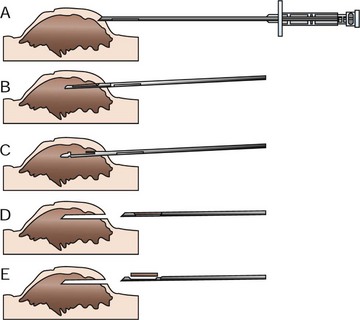
Biopsy Principles

9


Biopsy Principles


A biopsy refers to a procedure that obtains a tissue specimen for microscopic (i.e., histopathologic) analysis to establish a precise diagnosis. Histopathologic interpretation of tissue removed from a tumor is not infallible and is highly dependent on the quality of the biopsy sample submitted. Therefore it is important to understand basic principles of biopsy procurement and submission in order to obtain an accurate diagnosis. If the tissue diagnosis is incorrect, all subsequent steps in the treatment of the patient will also be incorrect.


Fine-needle aspiration cytology (FNAC) is a simple and rapid way to obtain information about a tumor and is often the first step in the diagnostic work-up (see Chapter 7). Results of FNAC help guide the diagnostic tests for staging. Studies have shown that FNAC is a reliable and useful method to guide further work-up when neoplasia is suspected or, in many cases, to help rule out neoplasia altogether.1,2 Nonetheless, FNAC gives only limited information and may be nondiagnostic or equivocal. Inflammation, necrosis, and hemorrhage may result in cytopathologic changes that do not accurately represent the underlying disease process. Histologic confirmation is therefore required for definitive diagnosis of neoplasia.


Many techniques are available for obtaining tissue specimens—ranging from needle-core techniques to complete excision. The choice of technique depends on the anatomic location of the tumor, the patient’s overall health, the suspected tumor type, and the clinician’s preference. Biopsy techniques can be grouped under one of two major categories: pretreatment biopsy (e.g., needle core biopsy, punch biopsy, wedge biopsy) or excisional biopsy. Pretreatment biopsy is performed in order to obtain additional information about the tumor prior to definitive treatment. Posttreatment (i.e., excisional) biopsy refers to the process of obtaining histopathologic information following surgical removal of the tumor. Excisional biopsy is best used to obtain a more complete picture of the disease process (e.g., histologic subtype, tumor grade, degree of invasion into regional vasculature and lymphatics) and provides an opportunity to evaluate completeness of excision. It is rarely the best first step in obtaining a tissue diagnosis. Although excisional biopsy is attractive to many clinicians because it allows for definitive treatment and diagnosis in one step, it is often used inappropriately in the management of a cancer patient, resulting in incomplete surgical margins. Incomplete surgical margins may result in local recurrence, the need for radiation therapy, or a wider, more extensive surgery. All of these sequelae represent compromise of the optimum treatment pathway for the patient and will involve more morbidity and expense than a properly performed first excision. The issue to be determined before surgery is: how aggressive should the surgery to remove the tumor be? It is intuitive that wide, ablative surgery (e.g., body wall resection) would be inappropriate for a simple lipoma. It also follows that marginal excision (“shell out”) is inappropriate for definitive treatment of an aggressive tumor, such as a soft tissue sarcoma. Thus thorough knowledge of the tumor type is imperative prior to attempting surgical excision. The best way to obtain this information is often via pretreatment biopsy.


Specific indications for pretreatment biopsy are as follows:


If any one of the listed criteria is met, a pretreatment biopsy should be pursued.


There are occasions when pretreatment biopsy would be contraindicated. These include cases when the type of treatment or extent of surgery would not be changed by knowing the tumor type (e.g., testicular mass, solitary splenic mass) or when the surgical procedure to obtain the biopsy is as risky as definitive removal (e.g., spinal cord biopsy). In these cases, the patient would best be served by excisional biopsy of the tumor if staging results support this choice.


Biopsy Methods


The more commonly used methods of tissue procurement are needle core biopsy, punch biopsy, incisional (wedge) biopsy, and excisional biopsy.


Needle Core Biopsy


Needle core biopsy utilizes various types of needle core instruments (e.g., Tru-Cut [Baxter General Healthcare, Deerfield, IL] or ABC needle [Kendall Sherwood-Davis & Geck, St. Louis, MO]) to obtain soft tissue (Figure 9-1). Most of these needles are manually operated, although spring and pneumatically powered needles are available as well. Specialized core instruments are used for bone biopsies and will be covered in Chapter 24. These instruments are generally 14-g in diameter and procure a piece of tissue that is about 1 mm wide and 1.0 to 1.5 cm long. In spite of this small sample size, the structural relationship of the tissue and tumor cells can usually be visualized by the pathologist. Virtually any accessible mass can be sampled by this method. It may be used for externally located lesions or for deeply seated lesions (e.g., in the kidney, liver, or prostate) with image-guidance via closed methods or at the time of open surgery.


The most common usage of the needle core biopsy is for externally palpable masses. Except for highly inflamed and necrotic cancers (especially in the oral cavity), in which incisional biopsy is preferred, most biopsies can be done on an outpatient basis with local anesthesia and sedation. The area to be biopsied should be clipped of hair and sterilely prepared. The skin or overlying tissue is prepared as for minor surgery. If the overlying tissue (usually skin and muscle) is intact, it is anesthetized using local anesthetic in the region that the biopsy needle will penetrate. Tumor tissue itself is very poorly innervated and generally does not require local anesthesia. The mass is then fixed in place with one hand or by an assistant. A small 1- to 2-mm stab incision is made in the overlying skin with a scalpel blade to allow insertion of the biopsy instrument. The stab incision is necessary to prevent dulling of the needle tip and allow better penetration into the underlying tissue. Through the same skin hole, several needle cores are removed from different sites to get a “cross-section” of tissue types within the mass. The stab incision can be sutured with a single interrupted suture. The tissue is gently removed from the instrument with a scalpel blade or hypodermic needle and placed in formalin. For smaller-gauge biopsy needle instruments, the tissue may be flushed off the needle with saline. Samples may be gently rolled on a glass slide for cytologic preparations before fixation. With experience, the operator can generally tell from the appearance of the core sample whether diagnostic material has been attained. Small, discontinuous bits of tissue and fluid within the trough will only rarely be diagnostic and usually imply the need for incisional biopsy. Soft tissue sarcomas in particular may not yield good tissue cores because of necrosis and fibrous septa that often permeate the mass. Cystic masses are also problematic.


Needle biopsy tracts are of minimal risk for local tumor seeding but should be removed en bloc with the tumor at subsequent resection. Therefore it is important to plan where the stab incision and needle biopsy tract are placed in order to make the subsequent excision simpler. Avoid excessive tunneling through uninvolved tissues by choosing the most direct path from the skin to the tumor to obtain a representative sample.


Many of these needles are “disposable” with plastic casings and therefore cannot be steam sterilized. It is not uncommon, however, for veterinary practices to resterilize these instruments (using ethylene oxide or hydrogen peroxide gas) and use them repeatedly until they become dull.


Needle core biopsy instruments are inexpensive and easy to use, and needle core biopsy procedures can be performed as outpatient procedures. They are generally more accurate than cytology but likely have lower accuracy than larger incisional or excisional biopsy, especially when a tumor is heterogeneous, inflamed, or cystic or contains a large amount of necrosis. It is important to understand that for a 5-cm diameter mass, one needle core biopsy sample represents less than 1% of the tumor tissue. The smaller the biopsy specimen obtained, the less representative it may be for the entire tumor.


Needle core biopsy can be performed with the aid of image-guidance (discussed in greater detail later). Utilization of image-guidance for needle core biopsy is helpful for obtaining tissue from deeply seated lesions. Ultrasound-, fluoroscopic-, and computed tomographic-assistance may be used to obtain samples from tumors located in areas where percutaneous biopsy would be risky or unlikely to yield a representative sample. In situations in which the lesion is located within a body cavity, the risk of tumor seeding from uncontrolled hemorrhage or fluid leakage as a result of image-guided biopsy must be taken into account when determining if image-guided needle core biopsy techniques hold an advantage over more direct access in a given circumstance.

Stay updated, free articles. Join our Telegram channel

Sep 22, 2016 | Posted by in SMALL ANIMAL | Comments Off on Biopsy Principles

Full access? Get Clinical Tree

Get Clinical Tree app for offline access