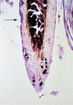
Bilaterally Symmetric Alopecia in Dogs

Chapter 39


Bilaterally Symmetric Alopecia in Dogs


Endocrine diseases are among the many causes of symmetric alopecia in the dog. In certain situations the cutaneous manifestations of endocrine disease provide some of the most obvious clinical signs. However, the clinician must consider a number of common and uncommon disorders when approaching the dog with symmetric alopecia (Box 39-1). As with most dermatologic disorders, signalment, historical information, and physical findings are the starting points to a diagnosis, followed by performance of appropriate laboratory tests and the collection of a dermatologic minimum database. This chapter provides a concise and rational guide for achieving a diagnosis when confronted with a canine patient with bilaterally symmetric alopecia. More details about these specific conditions can be found in this section and in Section V of this textbook.


Pruritic Alopecia


By definition, alopecia means hair loss. It must be determined if the dog was pruritic and created the alopecia from trauma (e.g., biting, scratching, rubbing) or if the hair fell out spontaneously. Most endocrine and metabolic causes of alopecia are nonpruritic unless complicated by other lesions. For example, dogs with spontaneous hyperadrenocorticism may become pruritic if calcinosis cutis is present. Likewise, dogs with various metabolic disorders including hyperadrenocorticism may develop pruritus if secondary infection from bacteria or yeast occurs. The only pruritic endocrine disorder is hyperestrogenism. Pruritus in general is usually symmetric, and therefore trauma-induced alopecia may mimic metabolic causes of symmetric alopecia. Regardless of the cause, bilaterally symmetric alopecia is rarely identical on both sides. The presence of similar appearing lesions on both sides of the midline allows us to use this terminology.


The presence of pruritus leads to a list of differential diagnoses that include myriad allergic, parasitic, and infectious diseases. Historical evidence supports trauma-induced pruritus. A trichogram (sometimes referred to as a KOH prep) is an easy diagnostic procedure used to determine if the tips of the hairs are broken due to traumatic causes. Normal hairs should have a fine taper at the tip of the hairshaft, whereas broken hairs have blunted ends. Hairs are grasped at their base with hemostats or thumb forceps and removed with traction. They are then placed on a glass slide with a few drops of mineral oil or potassium hydroxide (KOH). Care should be taken to place the hairs in the same direction so that they can be examined from base to tip. The application of a cover slip aids in viewing. The hairs can be examined under the microscope using either 4× or 10× magnification objectives. Lowering the light condenser makes viewing easier by increasing the refractivity.


A KOH prep also can be used to identify other conditions. For example, in dermatophyte-infected hairs fungal spores (microconidia) may be visible. Affected hairs have a damaged cuticle, are asymmetric, and are usually much wider than normal hairs. The presence of macromelanosomes along with a damaged hair cuticle may assist in the diagnosis of color-dilution alopecia (Figure 39-1); however, the definitive diagnosis is based on clinical signs and histopathologic evaluation. Interestingly, large melanosomes may be seen in color-dilute breeds such as the weimaraner, but the cuticle is not damaged; therefore these breeds do not exhibit color-dilution alopecia (with rare exceptions).


If broken hairs are present due to pruritic causes, every effort should be made to identify and treat the underlying cause of the pruritus. Of note is the historical use of glucocorticoid medications because such treatment could potentially interfere with interpretation of prior and future laboratory results.


Clinical Evaluation


Historical information is important in all dermatologic cases. Questionnaire templates are available for clinical use in virtually every veterinary dermatologic textbook. These are an efficient way to collect the necessary data to help formulate a list of differential diagnoses. They also provide the client an opportunity to reflect on the history, which hopefully leads to a more accurate account of the clinical problem. The signalment alone may provide important data and should not be overlooked. Many endocrine skin disorders can be considered more likely based on the age of onset. Likewise, many breeds are overrepresented for certain endocrine causes of alopecia.


Atypical coloration, such as a “blue” or “fawn” Doberman pinscher, may predispose the dog to color dilution alopecia at a later age. Although the Doberman is one of the most common breeds associated with this disorder, the Chihuahua, chow-chow, and Yorkshire terrier have increased prevalence. Any dog that exhibits a dilution pattern may develop color dilution alopecia. As puppies, affected dogs have a normal hair coat (other than color) and they may develop alopecia as the adult hairs replace the puppy coat. It is impossible to predict the age of onset or severity of the alopecia in color-dilute situations.


The sex status of the dog is also important when considering hyperestrogenism. Sertoli cell tumor is a testicular neoplasm more common in male dogs with retained testicles. Female dogs may develop postpartum hair defluxion. Lastly, traction alopecia may occur from the exuberant use of rubber bands or hair clips on some canine breeds; therefore the grooming and maintenance history is helpful in making this diagnosis.


A thorough investigation of all topical and systemic medications is required. Topical glucocorticoid medications can lead to local or systemic clinical signs such as skin atrophy, alopecia or follicular atrophy, changes in skin pigmentation, calcinosis cutis, and the development of secondary infections or demodicosis. Topical glucocorticoid medications have been responsible for inducing some spectacular symmetric alopecias and should not be ignored when investigating the underlying cause of the problem. Topical steroids also can have a profound influence on serum biochemical test results, especially serum alkaline phosphatase. Other commonly used medications such as ketoconazole may not directly lead to the development of symmetric alopecia but may influence laboratory results.

< div class='tao-gold-member'>

Stay updated, free articles. Join our Telegram channel

Jul 18, 2016 | Posted by in PHARMACOLOGY, TOXICOLOGY & THERAPEUTICS | Comments Off on Bilaterally Symmetric Alopecia in Dogs

Full access? Get Clinical Tree

Get Clinical Tree app for offline access