CHAPTER | 3 Bacterial Skin Diseases
Pyotraumatic Dermatitis (acute moist dermatitis, hot spots)
Features
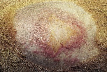
Pyotraumatic dermatitis is an acute and rapidly developing surface bacterial skin infection that occurs secondary to self-inflicted trauma. A lesion is created when the patient licks, chews, scratches, or rubs a focal area on its body in response to a pruritic or painful stimulus (Box 3-1). This usually is a seasonal problem that becomes more common when the weather is hot and humid. Fleas are the most common initiating stimulus. Pyotraumatic dermatitis is common in dogs, especially in thick-coated, long-haired breeds. It is rarely seen in cats.
Treatment and Prognosis
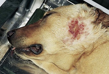
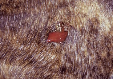
FIGURE 3-1 Pyotraumatic Dermatitis.
This moist, erosive lesion on the base of the ear is characteristic of a hot spot.
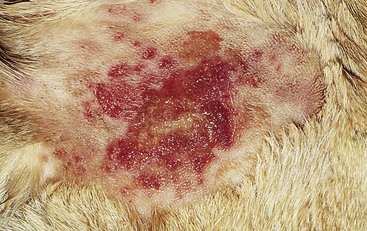
FIGURE 3-2 Pyotraumatic Dermatitis.
Close-up of the dog in Figure 3-1. The moist, erosive surface of the lesion is apparent. The papular perimeter suggests an expanding superficial pyoderma.
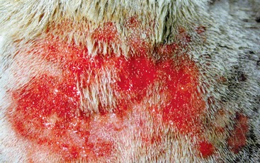
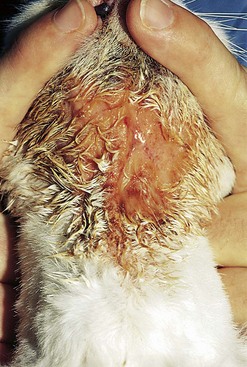
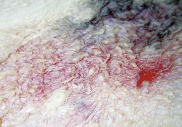
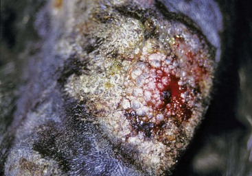
FIGURE 3-3 Pyotraumatic Dermatitis.
Close-up of a hot spot demonstrating the erosive lesion with a moist serous exudate.
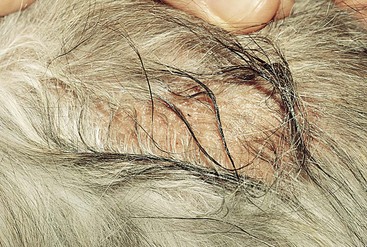
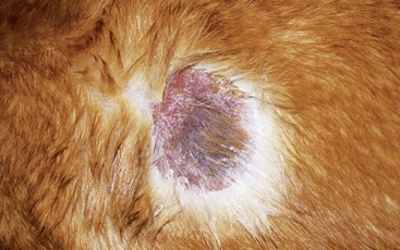
FIGURE 3-5 Pyotraumatic Dermatitis.
This moist lesion developed acutely on the dorsum of this flea-allergic cat.
Impetigo (superficial pustular dermatitis)
Diagnosis
Treatment and Prognosis
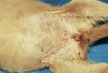
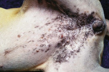
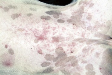
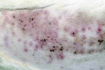
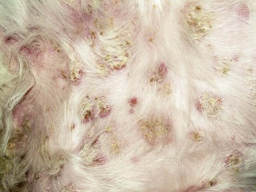
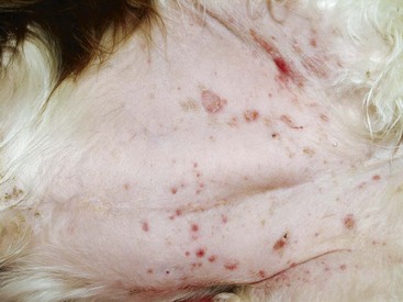
FIGURE 3-7 Impetigo.
Numerous superficial pustules and crusts on the abdomen of this puppy are typical of this disease.
Superficial Pyoderma (superficial bacterial folliculitis)
Features
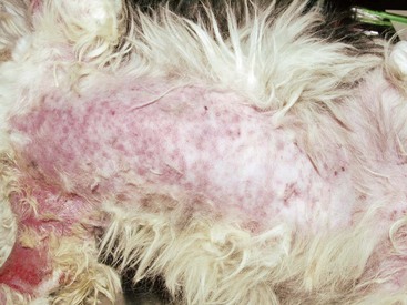
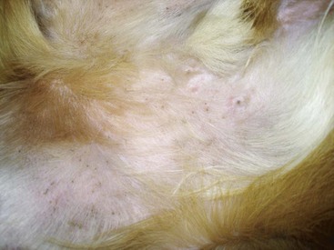
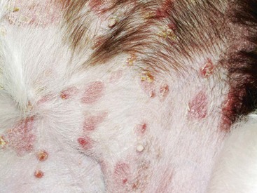
Superficial pyoderma is a superficial bacterial infection involving hair follicles and the adjacent epidermis. The infection is almost always secondary to an underlying cause; allergies and endocrine disease are the most common causes (Box 3-3). Superficial pyoderma is one of the most common skin diseases in dogs but is rare in cats.
Top Differentials
Differentials include demodicosis, dermatophytosis, scabies, and autoimmune skin diseases.
Top Diagnosis
Top Treatment and Prognosis
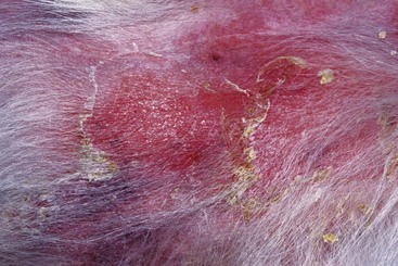
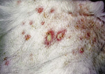
FIGURE 3-14 Superficial Pyoderma.
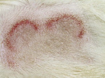
Close-up of the dog in Figure 3-13. Erythematous dermatitis with epidermal collarettes formation is apparent.
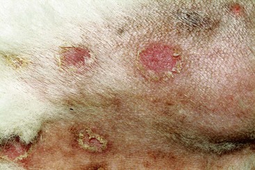
FIGURE 3-15 Superficial Pyoderma.
More typical epidermal collarettes in a dog with resolving pyoderma.
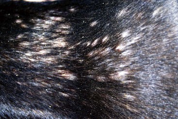
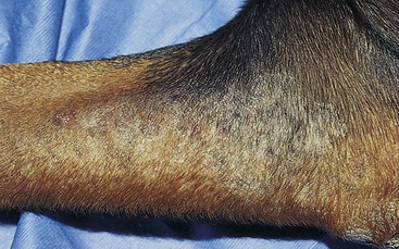
FIGURE 3-17 Superficial Pyoderma.
The moth-eaten alopecia is typical of pyoderma in short-coated breeds.
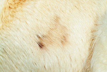
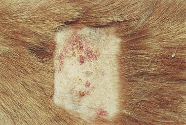
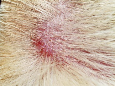
FIGURE 3-28 Superficial Pyoderma.
Focal area of alopecia caused by folliculitis in an allergic dog. Cutaneous cytology is necessary.
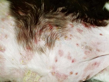
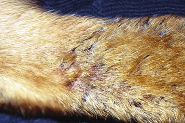
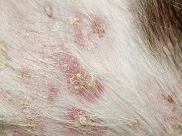
FIGURE 3-30 Superficial Pyoderma.
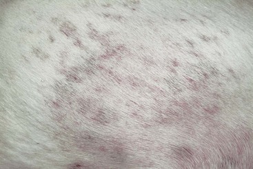
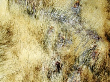
Multiple papules, crusts, and epidermal collarettes in a dog with hypothyroidism.
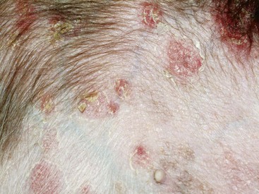
FIGURE 3-31 Superficial Pyoderma.
Close-up of the dog in Figure 3-30. Papular rash with crusting is apparent.
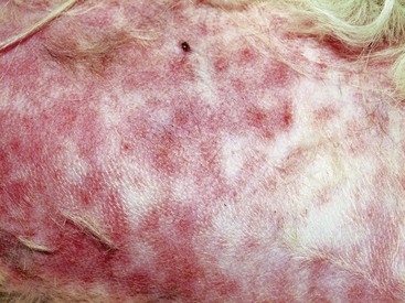
FIGURE 3-35 Superficial Pyoderma.
Same dog as in Figure 3-34. Erythematous macular lesions without a papular rash are apparent.
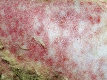
FIGURE 3-36 Superficial Pyoderma.
Same dog as in Figure 3-34. Erythematous macular lesions without a papular rash are apparent.
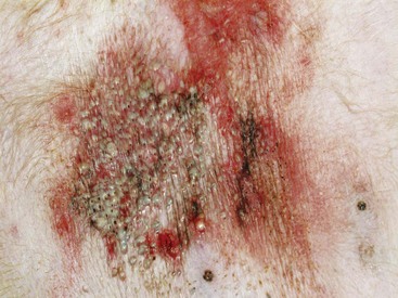
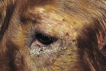
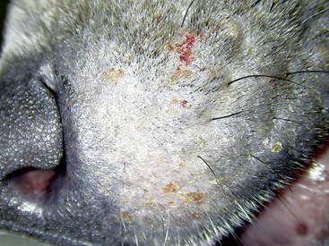
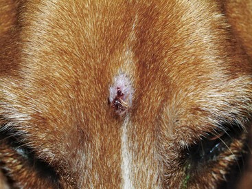
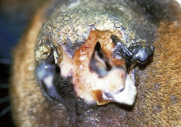
FIGURE 3-40 Superficial Pyoderma.
Severe inflammation caused by secondary bacterial infection. Comedones and pustules are visible.
Deep Pyoderma
Features
Deep pyoderma is a surface or follicular bacterial infection that breaks through hair follicles to produce furunculosis and cellulitis. Its development is often preceded by a history of chronic superficial skin disease, and it is almost always associated with some predisposing factor (see Box 3-3). Deep pyoderma is common in dogs and rare in cats.


