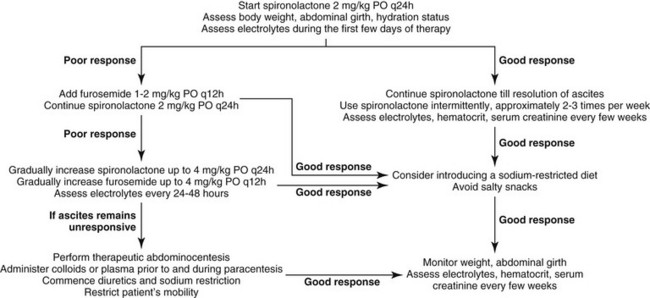
Chapter 144 Ascites and Hepatic Encephalopathy Therapy for Liver Disease Nick Bexfield, Cambridge, Great Britain Ascites Pathophysiology Ascites is the accumulation of fluid within the peritoneal cavity, and in liver disease this is usually a result of portal hypertension (PH). Hypoalbuminemia also can contribute to ascites formation in animals with reduced hepatic function, although it is unusual for hypoalbuminemia alone to cause ascites. PH, the sustained increase in blood pressure in the portal system, results from an increased intrahepatic resistance combined with increased portal blood flow. Increased intrahepatic resistance results from architectural distortion (fibrous tissue, regenerative nodules), sinusoidal endothelial dysfunction leading to impaired intrahepatic sinusoidal relaxation, and intrahepatic vascular shunts. With the development of PH, fluid is driven into the interstitial space, and when the capacity of the regional lymphatics is overwhelmed, ascites develops. The development of ascites is worsened by the splanchnic vasodilation that accompanies PH. This vasodilation results in pooling of blood in the abdomen, leading to a decrease in circulating volume, and when ionotrophic and chronotropic compensation fails, systemic hypotension results. This cumulates in activation of the renin-angiotensin-aldosterone system (RAAS), and volume expansion, which further increases hydrostatic pressure in the portal vasculature. PH is seen most commonly in dogs with chronic liver disease, although it occasionally occurs with acute liver disease; therefore the most common cause in dogs is chronic hepatitis progressing to cirrhosis. The presence of ascites is a poor prognostic indicator in dogs with chronic hepatitis, with a survival from the time of diagnosis of 0.4 months, compared to 24.3 months for nonascitic dogs. Because cats rarely get advanced liver fibrosis or cirrhosis, the development of PH and therefore ascites is uncommon in this species. Therapy for Ascites Diuretics Despite PH, ascitic animals actually have systemic hypotension and increased renal sodium retention because of reduced glomerular filtration rate and increased activation of the RAAS. Activation of the RAAS results in the release of aldosterone and increased sodium retention in the distal renal tubules. The aldosterone antagonist spironolactone is the diuretic of choice for the treatment of hepatogenic ascites. Spironolactone competes with aldosterone for its intracellular receptor sites, thus promoting sodium excretion and potassium retention in the renal tubules. Therapy with spironolactone at a dose of 2 mg/kg q24h PO is the initial therapy for animals with ascites resulting from PH (Figure 144-1). The dose of spironolactone can be increased gradually every few days to a maximum of 4 mg/kg q24h PO. Some animals may benefit from twice-daily therapy. Spironolactone can have a relatively slow onset of activity in humans, taking up to 14 days to cause diuresis; this also may occur in cats and dogs. In cases that are refractory to spironolactone, or when a more rapid resolution of ascites is required, furosemide (1 to 2 mg/kg q12h PO) also can be used. If there is no response to this dose of furosemide, it can be increased incrementally. Therapy with furosemide rather than spironolactone has been shown to precipitate more complications in humans with ascites, however. Importantly, serum electrolyte concentrations, especially sodium and potassium, should be monitored daily during the first few days of diuretic therapy, and every few weeks to months thereafter. Hypokalemia should be addressed as soon as possible as it can precipitate hepatic encephalopathy (HE) (see later). Body weight, abdominal girth (measure girth at level of the second lumbar vertebra with a tape measure), and hydration status as well as hematocrit and serum creatinine also should be monitored when on diuretic therapy. A safe loss of 0.5% to 1.0% body weight per day has been suggested for humans. Body weight reductions of 5% per day are dangerous and indicate the need for veterinary examination. When ascites has been mobilized adequately, intermittent use of diuretics is advised and guided by fluid reaccumulation. Administration of a diuretic two or three times per week is often sufficient to control fluid accumulation. Figure 144-1 A flow diagram describing the management of ascites in patients with liver disease. Abdominocentesis Abdominocentesis should be avoided if possible because the removal of large volumes of relatively protein-rich fluid can cause significant hypoalbuminemia, potentially leading to worsened fluid re-formation. Hypoalbuminemia also may lead to protein catabolism and HE. Therapeutic abdominocentesis is indicated if significant ascites is impairing mobility or causing respiratory compromise and is refractory to medical management. Fluid removal has the beneficial effects of improving cardiac function and stimulation diuresis. These effects are due to reductions of intraabdominal pressure, which compromises venous return. Administration of colloids or plasma before and during abdominocentesis is also advisable; this helps to prevent hypovolemia and worsening hypoalbuminemia as fluid shifts back into the abdomen. An infusion of 10 ml/kg of hetastarch over 3 hours is required, with abdominocentesis performed after the first 30 minutes of the infusion. Fluid removal is completed over 30 to 60 minutes.< div class='tao-gold-member'> Only gold members can continue reading. Log In or Register a > to continue You may also needPortosystemic ShuntsPortal Vein Hypoplasia (Microvascular Dysplasia)Diagnostic Approach to Hepatobiliary DiseaseChronic Hepatitis TherapyAcute Liver FailureCopper Chelator TherapySystemic HypertensionPulmonary Hypertension Share this:Click to share on Twitter (Opens in new window)Click to share on Facebook (Opens in new window)Click to share on Google+ (Opens in new window) Related Tags: Kirks Current Veterinary Therapy XV Jul 18, 2016 | Posted by admin in PHARMACOLOGY, TOXICOLOGY & THERAPEUTICS | Comments Off on Ascites and Hepatic Encephalopathy Therapy for Liver Disease Full access? Get Clinical Tree