Fig. 37.1
(a) A schematic drawing of “in vivo cryotechnique ” (IVCT) for living mouse sciatic nerves under various stretching conditions. A sciatic nerve of an anesthetized mouse is exposed, and its thigh is manually stretched. Liquid isopentane -propane cryogen (−193 °C) precooled in liquid nitrogen is then directly poured over the sciatic nerve. (b–g) Light microscopic (b–e) and confocal laser scanning microscopic (f, g) images of mouse sciatic nerves with IVCT (b, c, f) or perfusion fixation and alcohol dehydration method (PF-AD; d, e, g), stained with HE (b, d) or immunostained for albumin (c, e). By reconstruction of the optical sections, optimal three-dimensional image s are obtained (3D; f, g). Blue, nerve shapes with autofluorescence under UV; Red, albumin immunolocalization . With IVCT, albumin is immunolocalized in blood vessel s and the endoneurium (black arrowheads in c, f). In the node of Ranvier (white arrowheads in c, f), it is not immunolocalized inside the axon. With PF-AD, the albumin immunolocalization is observed heterogeneously in the endoneurium (black arrowheads in e, g) and inside the axons of some nerve fiber s (blue arrowheads in e, g). Precise data are reported in the previous paper (Kamijo et al. [14])
37.3 Immunolocalization of Albumin in Sciatic Nerve Fibers with IVCT
IVCT and freeze-substitution fixation (FS) have the immunohistochemical merit of keeping soluble protein s of living animal organ s in paraffin -embedded tissue sections [5, 9]. The serum albumin distribution in sciatic nerve fiber s and the structures of nerve fibers could be examined by immunohistochemistry (Fig. 37.1b, c), and the three-dimensional (3D) image of the albumin immunolocalizations was demonstrated by confocal laser scanning microscopy (CLSM) (Fig. 37.1f). Using autofluorescence signals with ultraviolet (UV) emission, nerve contour shapes were easily demonstrated (blue in Fig. 37.1f). The albumin immunolocalization was recognized with Alexa Fluor 594-conjugated secondary antibody (red in Fig. 37.1f). The albumin was immunolocalized in blood vessel s and the endoneurium (black arrowheads in Fig. 37.1c, f). In the node of Ranvier (white arrowheads in Fig. 37.1c, f), it was not immunolocalized inside the axon.
In sciatic nerves with perfusion fixation and alcohol dehydration (PF-AD), albumin was heterogeneously immunolocalized in the endoneurium (black arrowheads in Fig. 37.1d, e, g) and also inside the axons of some nerve fiber s (blue arrowheads in Fig. 37.1 d, e, g).
37.4 Structures of SLIs in Stretched and Non-stretched Sciatic Nerves with IVCT
The structures of nerve fiber s under the non-stretched condition by HE staining were mostly cylindrical (Fig. 37.2a). A membrane skeletal protein , 4.1G, was immunolocalized in paranodes and SLIs (black arrows in Fig. 37.2b) [15].
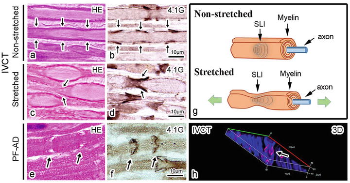
Fig. 37.2


Light microscopic (a–f) and confocal laser scanning microscopic (CLSM) (h) images of mouse sciatic nerves under non-stretched condition (a, b) and stretched condition (c, d, h) with IVCT or PF-AD (e, f), stained with HE (a, c, e) or immunostained for a membrane skeletal protein , 4.1G (b, d, f). Under the non-stretched condition, in cylindrical nerve fiber s (a), 4.1G is immunolocalized in Schmidt-Lanterman incisure s (SLIs: black arrows in b). Under the stretched condition, structures of nerve fibers show a beaded appearance (c). 4.1G-immunostained SLIs are observed in narrow areas and transitional regions from narrow to wide areas of the beaded nerve fibers (black arrows in d, h). By reconstruction of the optical sections with CLSM, three-dimensional image is obtained (3D; h). Images in (g) depict morphological changes of a nerve fiber including SLIs under non-stretched and stretched conditions. By PF-AD, structures of SLIs are disrupted (black arrows in e, f). Precise data are reported in the previous paper (Kamijo et al. [14])
Stay updated, free articles. Join our Telegram channel
Full access? Get Clinical Tree
