CHAPTER 194 Angular Limb Deformities
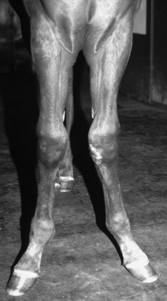
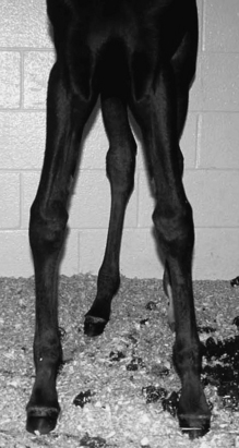
Angular limb deformities (ALDs) are defined as a deviation of the limb from the normal axis in the frontal plane. It is very common to have some degree of rotational deformity as well. Deviation of the limb laterally is termed valgus (Figure 194-1) and is most often accompanied by some degree of outward rotation of the limb, whereas deviation medially is defined as varus (Figure 194-2) and is often accompanied by inward or pigeon-toed rotation. ALDs may be congenital or acquired and usually affect the carpus, tarsus, and metacarpophalangeal or metatarsophalangeal joints.
ETIOLOGY
Congenital Angular Limb Deformity
Incomplete Ossification of Cuboidal Bones
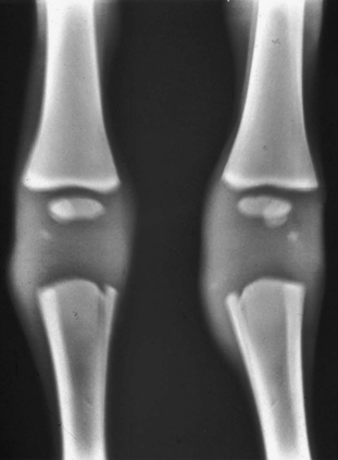
In an orthopedically normal foal, ossification of the cuboidal bones takes place during the last 2 months of gestation. In premature, dysmature, or twin foals, incomplete ossification of these bones is common (Figure 194-3). Most foals have mild carpal valgus at birth. This in itself is not usually a problem, but if the foal has incomplete ossification of the cuboidal bones, uneven joint loading may cause crushing of the hypoplastic cuboidal bones and can result in permanent ALD.

Figure 194-3 Anteroposterior view of the carpi of a foal with incomplete ossification of the cuboidal bones.
Acquired Angular Limb Deformity
Trauma or infection of the physis unevenly compresses the growth plate and reduces longitudinal growth. Excessive loading of the physis from overexertion or contralateral limb lameness causes the proliferative zone in the physis to be crushed, leading to microfractures, which ultimately result in early closure of the physis. This is also known as a Salter-Harris type-V fracture.


