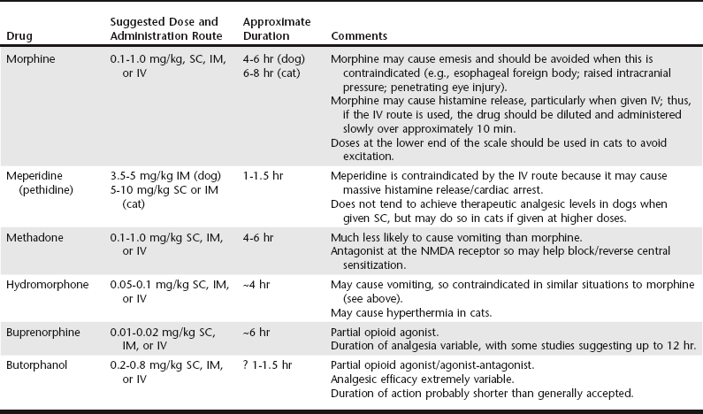
Anesthesia for the Critical Care Patient

Chapter 13


Anesthesia for the Critical Care Patient


Critically ill animals may require chemical restraint for a variety of procedures, ranging from minor interventions such as diagnostic imaging to major surgery. Although a clinician may be tempted to avoid anesthesia due to its inherent risks and instead perform complete procedures under sedation alone, deep sedation is likely to be a greater risk to the animal than “full” general anesthesia. Patients under deep sedation may be unable to maintain and protect their airway, may be breathing room air, and are rarely closely monitored during sedation. However, with general anesthesia the animal usually has an endotracheal tube in place, is receiving supplemental oxygen, and usually has one person dedicated to monitoring and maintaining physiologic function. This is not to say that sedative techniques should be avoided in critically ill animals, merely that they should only be used in situations in which the procedure can be performed under “light” sedation (often combined with local anesthesia) using drugs that are minimally depressant to the cardiovascular (CV) and respiratory systems. If these caveats cannot be met, general anesthesia is usually preferable. In all critical care patients undergoing any form of chemical restraint, secure intravenous access always should be available, and oxygen should be supplemented at every opportunity.


It is impossible to be prescriptive in terms of the anesthetic requirements of every critically ill animal, but, in general, this type of patient usually presents primarily with underlying dysfunction of the cardiovascular system, the respiratory system, or the central nervous system, although there is often multiorgan pathology.


Anesthesia for Patients with Cardiovascular Dysfunction


CV dysfunction may be related to volume depletion, vascular dysfunction, heart rhythm disturbances, advanced heart disease, or overt congestive heart failure. Appropriately categorizing CV risk and dysfunction is an important aspect of sedation and anesthesia of these patients. Special consideration should be directed to the preanesthetic period, induction of anesthesia, and maintenance.


Specific Patient Groups


The main forms of CV dysfunction encountered in critical care patients relate to hypovolemia or to cardiac arrhythmias. Additionally, some older dogs are affected by advanced valvular heart disease or chronic heart failure. In cats cardiomyopathies (sometime occult) are relatively common.


Hypovolemic Patients


Hypovolemia may be present as a result of dehydration (e.g., protracted vomiting with intestinal obstruction) or blood loss. Virtually all sedative and anesthetic agents cause a degree of CV depression, so the use of these drugs in patients with depleted circulatory volume can result in a severe drop in arterial blood pressure (BP). Consequently, restoration of the circulating blood volume should be undertaken prior to any form of chemical restraint, although there may be an argument for “delayed resuscitation” in those animals with internal hemorrhage (see Chapter 1).


Even following adequate volume resuscitation in hypovolemic patients, it is wise to provide chemical restraint by choosing agents with the least CV-depressant effects and using the lowest doses possible, usually through coinduction and balanced anesthetic techniques (see sections on “Induction of Anesthesia” and “Maintenance of Anesthesia”).


Patients with Cardiac Arrhythmias


Cardiac arrhythmias are common in critical care patients. Although these may result from underlying cardiac disease, a number of extracardiac factors also may be responsible (e.g., hypoxemia, hypercapnia, electrolyte disorders). Regardless of etiology, the aim should be to restore normal cardiac rhythm before induction of anesthesia through treatment of underlying pathology, correction of exacerbating factors (e.g., hypokalemia), and judicious use (when necessary) of appropriate antiarrhythmic drugs (see Chapters 171 and 172). However, even with these measures, restoration of normal sinus rhythm may not always be possible, and consideration needs to be given to selection of an anesthetic technique that will not further exacerbate the arrhythmia.


The main aims in critically ill patients with preexisting cardiac arrhythmias are to avoid agents that are pro-arrhythmic (e.g., α2-adrenergic agonists, thiopental, halothane) and use balanced anesthetic techniques to minimize the dose of any one agent. Hypoxemia, hypercapnia, and hypotension should be avoided in these animals because they may significantly exacerbate any preexisting arrhythmia. Thus consideration should be paid to preoxygenation prior to induction of anesthesia, oxygen supplementation during the procedure (even if the procedure is performed solely under sedation), and possibly the use of intermittent positive pressure ventilation to avoid excessive hypercapnia, accepting that controlled ventilation may carry detrimental CV side effects (see the section on “Intermittent Positive Pressure Ventilation” later). Drugs that may be required to manage acute exacerbations of the arrhythmia (such as lidocaine) or extremes of heart rate (atropine or glycopyrrolate; esmolol or propranolol) should be readily available. Not only should these drugs be close at hand, but the appropriate volume to be administered calculated in advance. The electrocardiogram should be monitored before, during, and after the anesthetic periods.


Premedication/Sedation of Patients with CV Dysfunction


A significant proportion of critical care cases with CV dysfunction may not require sedative premedication, particularly those that are severely hypovolemic. In general, patients with any degree of hypovolemia should not be given acepromazine because it causes vasodilation and will drop BP. The α2-adrenergic agonists (xylazine, medetomidine, dexmedetomidine) also should be avoided because they profoundly decrease cardiac output by affecting afterload and heart rate. In contrast, both opioids and benzodiazepines are relatively cardiostable and are extremely useful when dealing with high-risk CV cases.


Although paradoxical excitement can occur when benzodiazepines alone are administered to healthy animals, this is much less likely when used in depressed, critically ill patients. Combining these drugs with an opioid also improves the degree of sedation and limits the likelihood of excitation. The two benzodiazepines commonly used in small animals are diazepam and midazolam. Diazepam is best administered intravenously because it is poorly absorbed and relatively painful when given intramuscularly; midazolam is preferable if the intramuscular route is to be used. Although the latter is significantly more expensive than diazepam, it can be mixed in the same syringe with opioid drugs; this cannot be done with diazepam because precipitation may occur.


A variety of opioids (see Chapter 12) may be used in combination with the benzodiazepines, and the choice depends to a large extent on the procedure that the animal is undergoing. If there is preexisting moderate to severe pain, or if such pain is anticipated following the procedure, a full mu-agonist such as oxymorphone, hydromorphone, morphine, or methadone would be most appropriate because these can be titrated to the degree of pain. Partial agonists such as butorphanol or buprenorphine may be suitable alternatives but are less efficacious analgesics and should be limited to mild to moderate pain only. The full opioid agonists can also be antagonized (most commonly by naloxone) if the situation suddenly deteriorates, although this is uncommon if excessive doses are avoided. Table 13-1 lists suggested doses for each agent.


There is huge variability between individual patients in terms of dose requirements and analgesic response, and the doses/durations in Table 13-1 are intended only as a general guide.


Induction of Anesthesia


Most of the commonly used induction agents (thiopental, propofol, alfaxalone) can cause hypotension even in healthy patients, and in critical animals this can be profound. For this reason, these agents are not recommended by themselves for anesthetic induction in patients with limited CV reserve. Although combinations of ketamine/diazepam are considered relatively “cardiac-safe” in healthy animals (in that cardiac output and arterial BP are usually well maintained), the high preexisting (often maximal) sympathetic tone evident in most critically ill patients means that the animal may not be able to compensate for the direct negative inotropic effects of ketamine in the same manner as healthy animals; as a result, BP may fall to a similar extent to that of the previously discussed induction agents.


Etomidate is a nonbarbiturate sedative-hypnotic drug often used as an intravenous induction drug in human patients with CV disease because it has minimal effects on cardiac output and arterial BP. Although not licensed for veterinary use, it has been extensively used in dogs and cats with significant preexisting arrhythmias, cardiac pathology, or ongoing hypovolemia. This drug must be used with attention to potential adverse effects. Due to the high osmolarity of the solution, etomidate can cause pain on injection and thrombophlebitis. As a result, it is probably best administered into a free-flowing intravenous fluid line so it is suitably diluted before it enters the patient’s vein; this is probably more important in cats due to the small vein size. Etomidate can also cause myoclonus (twitching) at induction, as well as excitement and muscle movements during anesthetic recovery; this is much more common if the drug has not been preceded by a benzodiazepine or a potent opioid such as fentanyl. Additionally, etomidate causes suppression of cortisol production, so should only be administered as a single intravenous induction dose or acute hypoadrenocorticism may be observed. Some anesthesiologists recommend administering physiologic doses of glucocorticoids on the day that a patient receives etomidate.


With all the aforementioned anesthetic induction agents, it is essential to minimize the dose used when administered to patients with unstable CV status. This can be achieved using a coinduction technique, which implies the use of one or more additional drugs alongside the main hypnotic agent. The coinduction drug(s) used should be capable of reducing the dose of hypnotic required to produce unconsciousness, while at the same time having minimal CV-depressant effects of their own. The most common coinduction agents used are the potent, short-acting opioids, such as fentanyl and alfentanil, and the benzodiazepines, diazepam and midazolam. For coinduction, one of these opioids or benzodiazepines (or a combination) is administered intravenously immediately before the chosen hypnotic agent, resulting in a reduced dose requirement for the latter and reduced CV depression.


As an alternative, and particularly for those animals with severe CV compromise in which even a small dose of hypnotic drug may not be tolerated, a combination benzodiazepine/opioid induction protocol can be very useful. Because both groups of drugs are relatively cardiostable, they can be given in fairly high doses to achieve induction of anesthesia. The most common technique utilizes a combination of fentanyl and diazepam: a dose of 10 µg/kg fentanyl and a dose of 0.5 mg/kg diazepam are drawn up into separate syringes. After a 3- to 5-minute period of preoxygenation by facemask, half the fentanyl (5 µg/kg) is administered intravenously, followed by half of the diazepam (0.25 mg/kg). Adequate time must be allowed for the drugs to reach peak effect, since this technique results in slower induction than traditional intravenous induction techniques. In addition, the appearance of animals following benzodiazepine/opioid induction is different to that following “classical” anesthetic induction; these patients may occasionally remain in sternal recumbency and often appear quite awake, with centrally positioned glazed eyes. However, even in this state many still tolerate intubation of the trachea. If anesthesia is inadequate for endotracheal intubation, further increments of the two drugs are delivered until unconsciousness is achieved. On occasion, particularly if the animal was not significantly depressed prior to induction of anesthesia, inadequate hypnosis may not be achieved after the full doses of diazepam and fentanyl have been administered; unconsciousness can then be produced with an intravenous hypnotic such as propofol or alfaxalone, but only very small doses of these agents are required due to the anesthetic sparing effects of both fentanyl and diazepam. High doses of fentanyl can result in bradycardia; this can usually be avoided if the drug is given very slowly over several minutes, but it is wise to have an antimuscarinic (atropine or glycopyrrolate) drawn up and ready to administer, if necessary. Alternatively, some authorities recommend pretreatment with an antimuscarinic if a benzodiazepine/opioid induction technique is to be used, although this may in itself lead to some CV instability due to the likely transient tachycardia.

< div class='tao-gold-member'>

Stay updated, free articles. Join our Telegram channel

Jul 18, 2016 | Posted by in PHARMACOLOGY, TOXICOLOGY & THERAPEUTICS | Comments Off on Anesthesia for the Critical Care Patient

Full access? Get Clinical Tree

Get Clinical Tree app for offline access