chapter 21 Alternative Imaging Technologies
Upon completion of this chapter, the reader should do the following:
Acoustic impedance: Relationship between density or stiffness of tissue and the velocity of sound within the tissue. Differences in acoustic impedance of adjacent tissues determine the intensity of reflected sound.
Acoustic shadow: Ultrasound artifact. Echo-free zone created distal to the imaged organ when sound waves hit a highly reflective tissue that prevents sound from being transmitted to greater depths.
Anechoic: No echoes are detected, and the area is black. Typically associated with fluid-filled structures such as the urinary bladder.
Attenuation: Reduced intensity of radiation caused by absorption or scattering, or both, during passage through tissue. Sound is also attenuated as it passes through tissue and the intensity is reduced.
B-mode (brightness-mode) ultrasonography: Intensity of returning echoes is expressed as brightness in the display.
Computed tomography (CT) number: Number converted to gray scale in the final image, which represents the attenuation of the x-ray beam in tissue within a voxel. The number is also referred to as a Hounsfield number, named for the inventor of CT scanning.
Curie (Ci): A unit of activity (3.7 × 10¹⁰ disintegrations per second).
Distant enhancement: Ultrasound artifact. Increased sound intensity beyond a fluid-filled, anechoic area, created by absence of attenuation of the sound beam as it passes through the fluid.
Doppler shift: Difference between transmitted and received sound frequencies. The greater the Doppler shift, the greater the flow velocity.
Echogenicity: Intensity of reflected echoes.
Half-life (t½): Time in which the initial activity of a radionuclide is reduced to one half. Biologic half-life includes excretion, as well as the characteristic half-life of the isotope.
Hyperechoic: Echoes produced are brighter than in surrounding tissue.
Hypoechoic: A few echoes are detected, and the area is low-level gray compared with adjacent tissues. Usually seen with solid homogeneous tissues or complex fluid containing cells such as blood.
Labeled compound: A compound whose molecule is tagged with a radionuclide.
Linear array probe: Ultrasound probe containing multiple in-line transducers that create a rectangular-shaped image.
Long-axis view: Echocardiographic image showing the heart from base to apex in a longitudinal or sagittal plane.
M-mode (motion-mode) ultrasonography: Information is displayed as depth versus time on a graph. Used for echocardiography.
Pixels (picture elements): Tiny squares making up the image matrix; represent voxels.
Radiopharmaceutical: A radioactive drug that can be administered for diagnostic or therapeutic purposes.
Sector probe: Ultrasound probe with multiple rotating or oscillating transducers that produce a wedge-shaped image.
Short-axis view: Echocardiographic image showing the heart in transverse plane.
Target organ: The organ intended to be imaged and expected to receive the greatest concentration of administered radioactivity.
Voxel (volume element): Three-dimensional box represented on an image matrix by the two-dimensional pixel.
ULTRASONOGRAPHY
Ultrasonography has been an important imaging modality in veterinary medicine since the 1980s. Ultrasound can provide information about organ architecture independent of organ function. It is especially helpful in debilitated or young patients, in which the contrast agents used in special procedures or exploratory surgery may be contraindicated. Ultrasonographic findings are not necessarily specific for histopathologic diagnoses. However, the ability to distinguish solid masses from those containing fluid and to determine the distribution of lesions in organs allows the sonographer to focus differential diagnoses and to formulate management plans.
Technical Aspects
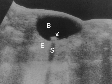
The ultrasound beam is created by a piezoelectric crystal that oscillates at several million Hertz per second (MHz) within a transducer (probe). When the sound wave interacts with tissues in the body, it is reflected, and the echo is received by the transducer. The received impulse is converted to an electronic signal and processed through a computer to become part of a composite of signals that make up the final image of the organ. Differences among organs are identified on a survey radiograph because of the different x-ray attenuating properties that tissues have. With ultrasound, returning signals have different intensities because tissues have different acoustic properties or acoustic impedance. Elasticity of the tissue determines the way sound interacts with the tissue: reflection, transmission, or refraction (Fig. 21-1). Air scatters sound. Water transmits sound with little attenuation or reflection. This lack of attenuation creates distant enhancement, an ultrasound artifact that indicates the presence of fluid. Minerals and metals are highly reflective. Sound cannot penetrate bone. This results in acoustic shadowing, which is a lack of echoes beyond the reflecting object. The echogenicity of tissues is an indication of the liquid or solid composition of the tissue. Anechoic tissues reflect few, if any, echoes. A full urinary bladder is anechoic. Hypoechoic tissues reflect few echoes. The medullary papillae of the kidney are hypoechoic. Hyperechoic tissues reflect bright white echoes. A bladder stone is hyperechoic.
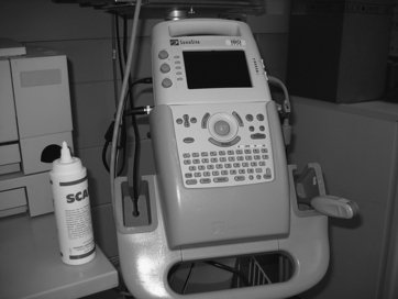
Ultrasound machines display images in real time. Sector probes or linear array probes are applicable for small and large animals; typically, 5- and 7.5-MHz transducers are used. A large animal practice may also require lower-frequency probes such as 3- or 2.5-MHz (Fig. 21-2). The frequency of the probe is tailored to the size of the animal and to the depth of the organ or area to be imaged. A higher-frequency probe provides better resolution and detail. However, the depth to which the sound can penetrate is limited to areas closer to the surface. A 7.5-MHz transducer is effective in cats and small dogs or for equine reproductive and tendon work. A 5-MHz transducer is used for medium- to large-breed dogs and for equine reproductive scanning. To penetrate at greater depths, a lower-frequency transducer is used. The detail in the image, however, is not as sharp.
Clinical Applications
Echocardiography
M-mode (motion-mode) and two-dimensional B-mode (brightness-mode) echocardiography are used to evaluate cardiac disease (myocardial and valvular disease, as well as congenital anomalies). To perform an echocardiogram, there is no specific preparation. Restraint of the patient is necessary to protect personnel and equipment, and sedation is rarely necessary. An area of chest wall over the heart is clipped. Acoustic gel is applied to conduct sound from the transducer to the thoracic wall. The left and right cardiac windows, located just caudal to the elbow, are used. The forelimb on the side to be imaged is pulled forward to permit directional freedom of the transducer and to prevent superimposition of bone or muscle over the window (Fig. 21-3). If the ribs are so close together that the transducer head cannot make contact with the chest wall, or if the available window is too narrow (which is often the case in cats), a pillow, rolled-up towel, or sponge wedge may be placed beneath the patient to spread the ribs. In the normal approach, the transducer is directed downward toward the chest wall, which is on the upper side of the patient. The patient may also be approached from the dependent side by directing the transducer upward from beneath table level. Imaging of a patient in sternal or in standing position is also an option, especially in large-breed dogs. Echocardiography is especially difficult in deep-chested breeds such as the Irish wolfhound or the Borzoi and in barrel-chested breeds such as the bulldog because of their conformation.
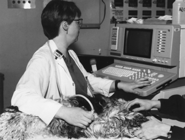
Figure 21-3 Echocardiography performed on a dog showing positioning for the right parasternal approach to the heart.
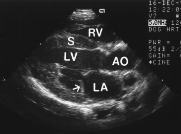
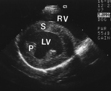
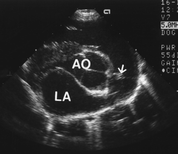
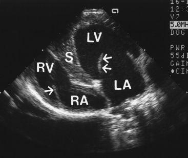
Two-dimensional echocardiography improves understanding of cardiac and regional mediastinal anatomy. Several standard views are obtained in both the long-axis and the short-axis directions (Figs. 21-4 through 21-7). Long-axis scans should include the left atrium, mitral valve, interventricular septum, and left ventricular free wall. By slightly tipping the transducer, the aortic valve and aortic root can be seen. In cats the right atrium and right ventricle are difficult to see on the long-axis view when approached from the right parasternal position because the right side of the cat heart is so close to the thoracic wall, and this side of the heart is often outside of the focal zone of the transducer. In dogs the right heart chambers are usually seen on the long-axis view from this position. Short-axis scans should include structures from the base to the apex of the heart. The aortic valve, pulmonary trunk, pulmonic valve, and left atrium are seen at the base of the heart. The mitral valve is the next structure to be seen as the transducer is directed more toward the apex of the heart. Below the mitral valve level are the left ventricle, interventricular septum, and right ventricle. The four-chamber view shows the left and right atria, the mitral and tricuspid valves, and the right and left ventricles. This view is obtained from the left parasternal position.
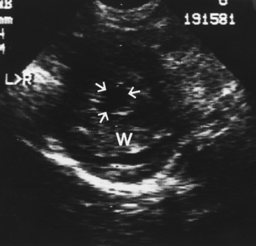
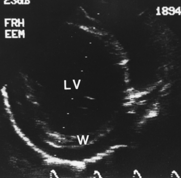
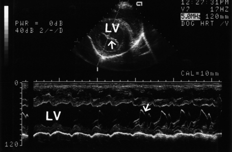
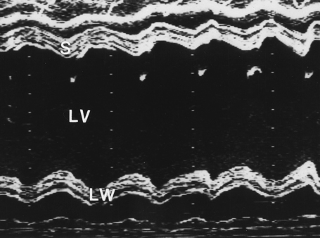
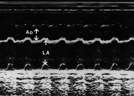
A quick overview of the anatomy and function of the heart allows a rapid assessment of functional compromise and detection of obvious chamber size abnormalities (Figs. 21-8 and 21-9). Two-dimensional scans are helpful in identifying cardiac abnormalities such as pleural and pericardial effusion, cardiac masses, and congenital anomalies such as defects in the interventricular septum. An M-mode examination is optimal to detect abnormal valvular motion such as fluttering, prolapse, or insufficient closure and to accurately measure chamber size and wall thickness. M-mode ultrasound displays cardiac wall and valvular movement as a graph over a period of time (Fig. 21-10). The graph represents the distance of structures from the transducer on the vertical axis and allows the investigator to measure the thickness of the interventricular septum, left ventricular and atrial chamber size, left ventricular wall thickness, and the aortic outflow track (Figs. 21-11 and 21-12). Aortic and mitral valvular motion and thickness can also be assessed. The right thoracic wall is approached to obtain the M-mode views used for measurements. Cardiac function is determined from the dimensions of the left ventricular lumen in systole and diastole to calculate fractional shortening (contractility).
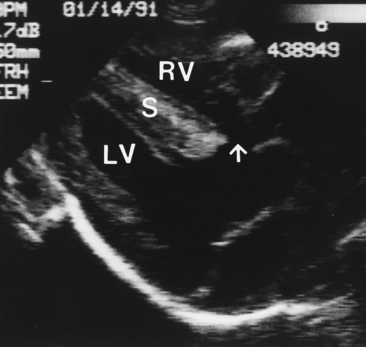
Horses are scanned in a standing position, usually confined in stocks. Indications for echocardiography of the horse are congenital heart disease and acquired valvular disease. Ventricular septal defect (VSD) is the most commonly diagnosed lesion (Fig. 21-13). Acquired valvular disease is commonly seen in middle-aged to older horses. Myocardial disease and pericardial effusion are uncommon in the horse.
Stay updated, free articles. Join our Telegram channel

Full access? Get Clinical Tree


